|
||
|
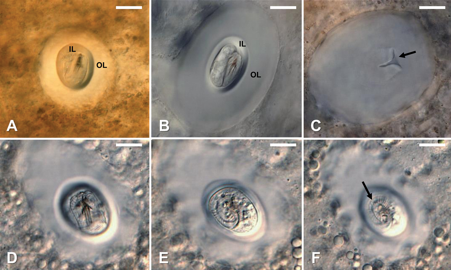
Gordius terrestris sp. nov., cysts, light photomicrographs A–B fully formed cysts in experimentally infected Physa acuta snails; note the folded larva surrounded by a clear cyst wall of unknown composition with a distinct inner layer (IL) and outer layer (OL) C remaining cyst wall after the folded larvae was extruded under coverslip pressure. Note the opening where the larvae emerged (arrow) D–F different focal planes showing the distinct larvae folding pattern; note the location of the terminal spine (arrow) in F and that the larva folds twice within the fully formed cyst. Scale bars: 20 µm (A–F). |