|
||
|
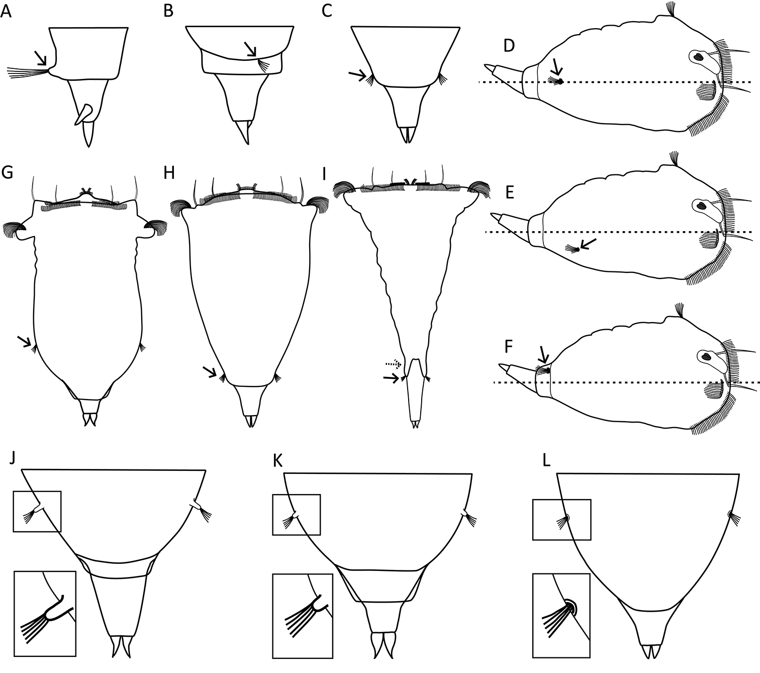
Location and morphology of the lateral antennae. A–C Number and size of the lateral antenna(e) (arrow) A single, enlarged left lateral antenna (S. hutchingsi) B single, right lateral antenna (S. tamara) C lateral antennae paired, symmetrical, and of normal size (S. tremula) D–F Location of the lateral antenna(e) (arrows) relative to the median transversal axis (dashed line) (lateral habitus is presented as a stylized drawing that is species independent) D directly lateral (e.g., S. tremula) E ventrolateral (e.g., S. oblonga) F mid-dorsal, single antenna slightly displaced to the right of the body axis (e.g., S. tamara) G–I Location of the lateral antennae relative to the longitudinal axis (arrows) G in the posterior third of the trunk region (S. oblonga) H in the caudal-most trunk region at or near the base of the foot (S. tremula) I on lateral lobes (dashed arrow) caudally to the cloaca and in the proximal third of the foot (S. grimpei) J–L The base of the lateral antennae (detail in inset) J surrounded by a tubular epidermal fold (S. johanseni) K surrounded by a papillary epidermal fold (S. oblonga) L surrounded by a low epidermal fold (S. pectinata). Drawings modified from: B Smirnov (1933) and Friedrich and De Smet (2000) J Harring (1921). |