|
||
|
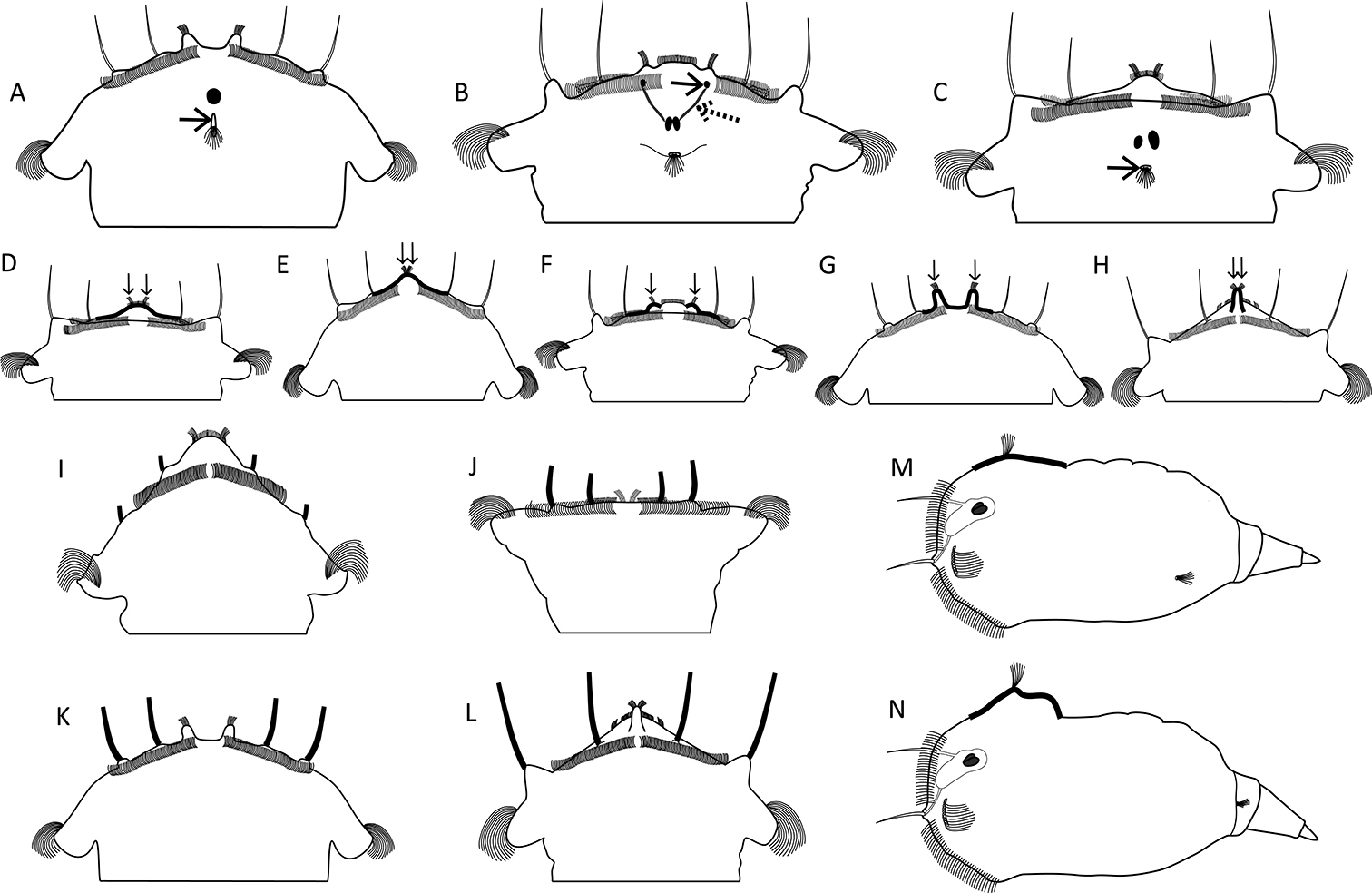
Sensory system. A–C Head region showing the cerebral eye, pigment granules and the opening of the dorsal antenna A cerebral eye single, dorsal antenna opening slit-shaped (arrow; S. pectinata) B two partially fused cerebral eyes, frontal aggregations (arrow) and streams (dashed arrow) of pigment granules present (S. triophthalma) C cerebral eyes distinctly separated, dorsal antenna opening round (arrow; S. oblonga) D–H Morphology of the apical receptors (thickened lines, arrows) D receptors slightly separated, situated on a slight elevation centrally on the apical field (S. oblonga) E receptors incompletely separated, situated on a strong elevation centrally on the apical field (S. grandis) F receptors distinctly separated, each situated on a bulge (S. triophthalma) G receptors distinctly separated, each situated on a strong tentacle-like elevation (S. pectinata) H receptors incompletely separated, situated on a single tubular elevation (S. vorax) I–L Lengths of the lateral and dorsolateral styles (thickened lines) I minute (S. squamadigitata) J short (S. grimpei) K medium (S. pectinata) L long (S. vorax) M, N Elevation underlying the dorsal antenna (thickened lines) M not elevated to slightly elevated (S. oblonga) N distinct prominence (S. tremuloida). Drawings modified from: I De Smet (2006). |