|
||
|
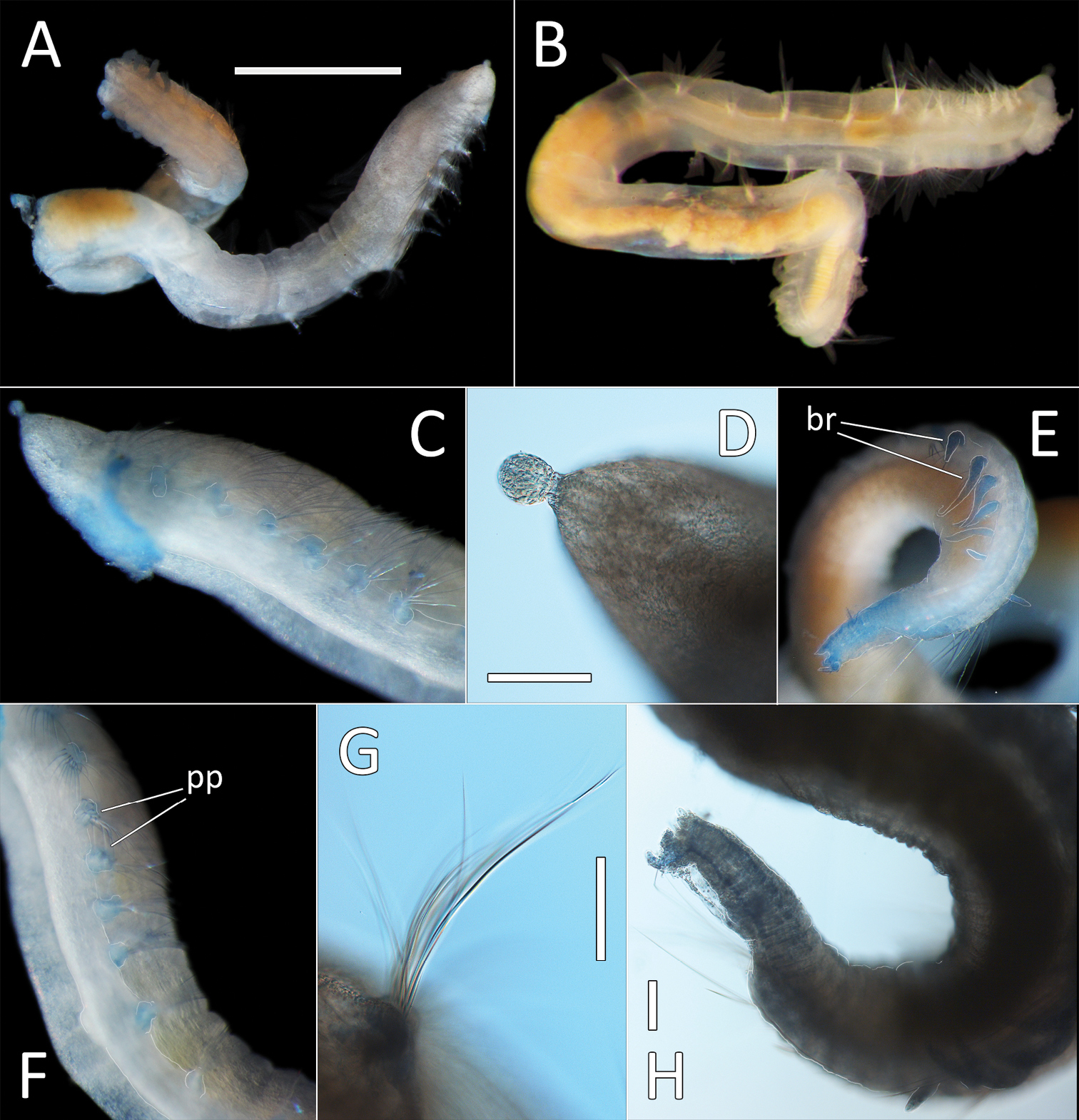
Ammotrypanella sp. NHM_1653 (specimen NHM_1653). A Lab image, whole specimen (faded stain B Live image, whole specimen C Lab image, anterior (faded stain) D Lab image, detail of palpode E Lab image, posterior (faded stain, br = branchiae) F Lab image, anterior-midbody (faded stain, pp = parapodia) G Lab image, detail of capillary chaetae H Lab image, detail of posterior and anal tube. Morphological features in plates C, E, F, G have been outlined with a very fine white to improve clarity of those features. Scale bars: 1 mm (A); 100 μm (D, G, H). |