|
||
|
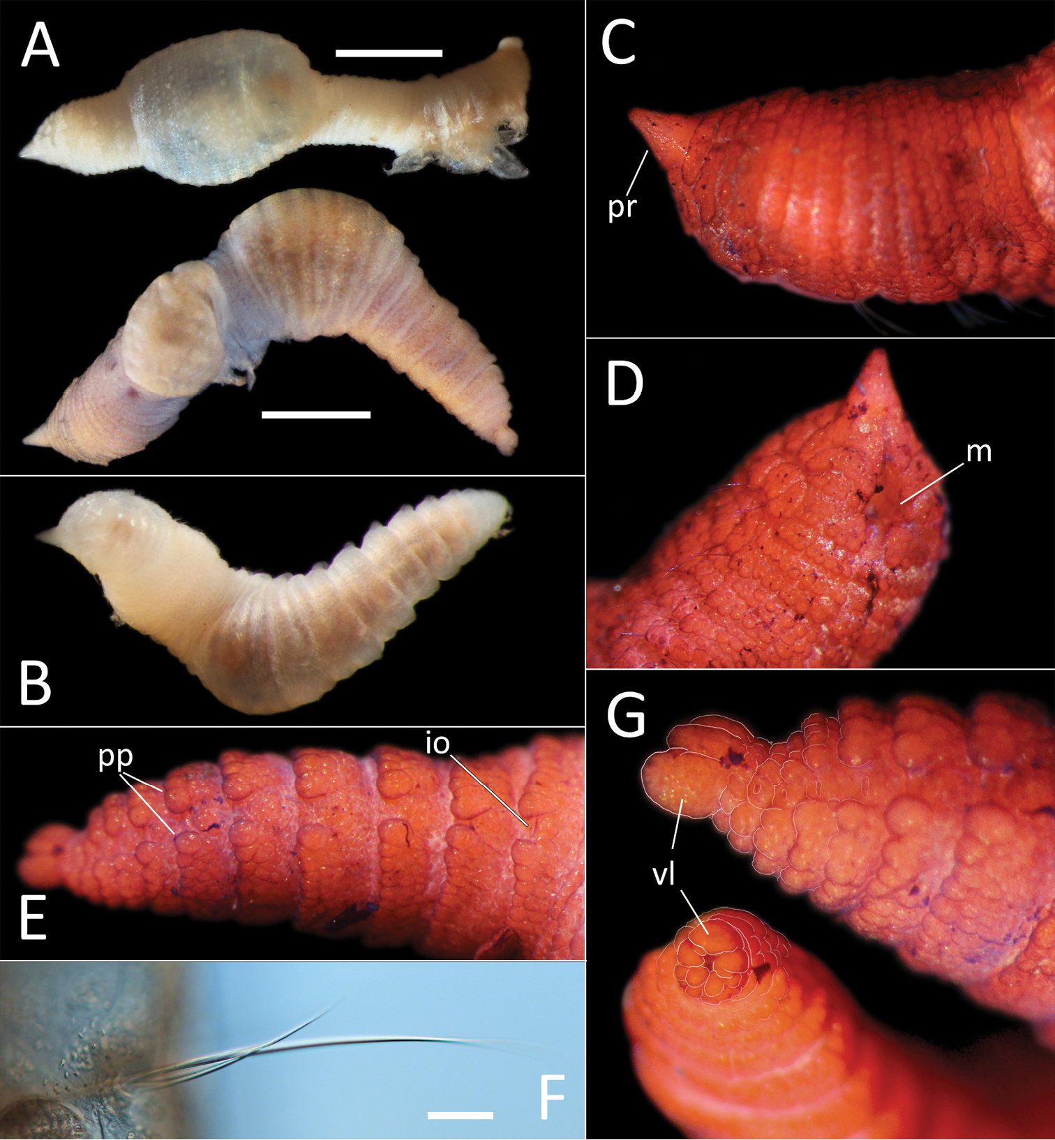
Travisia sp. NHM_1244. A Lab images, whole specimens (specimen NHM_1244, unstained [top], specimen NHM_1863, faded stain [bottom]) B Live image, whole specimen (specimen NHM_1863) C Lab image, dorsal anterior (specimen NHM_1863, stained, pr = prostomium) D Lab image, ventral anterior, (stained) (specimen NHM_1863, m = mouth) E Lab image, lateral posterior (specimen NHM_1863, stained, pp = parapodia, io = interramal organs) F Lab image, detail of capillary chaetae (specimen NHM_1244) G Lab image, pygidium, distal view (lower left) and lateral view (upper right), with pygidial features outlined in a fine white line (specimen NHM_1863, stained, vl = ventral lobe). Scale bars: 1 mm (A); 50 μm (F). |