|
||
|
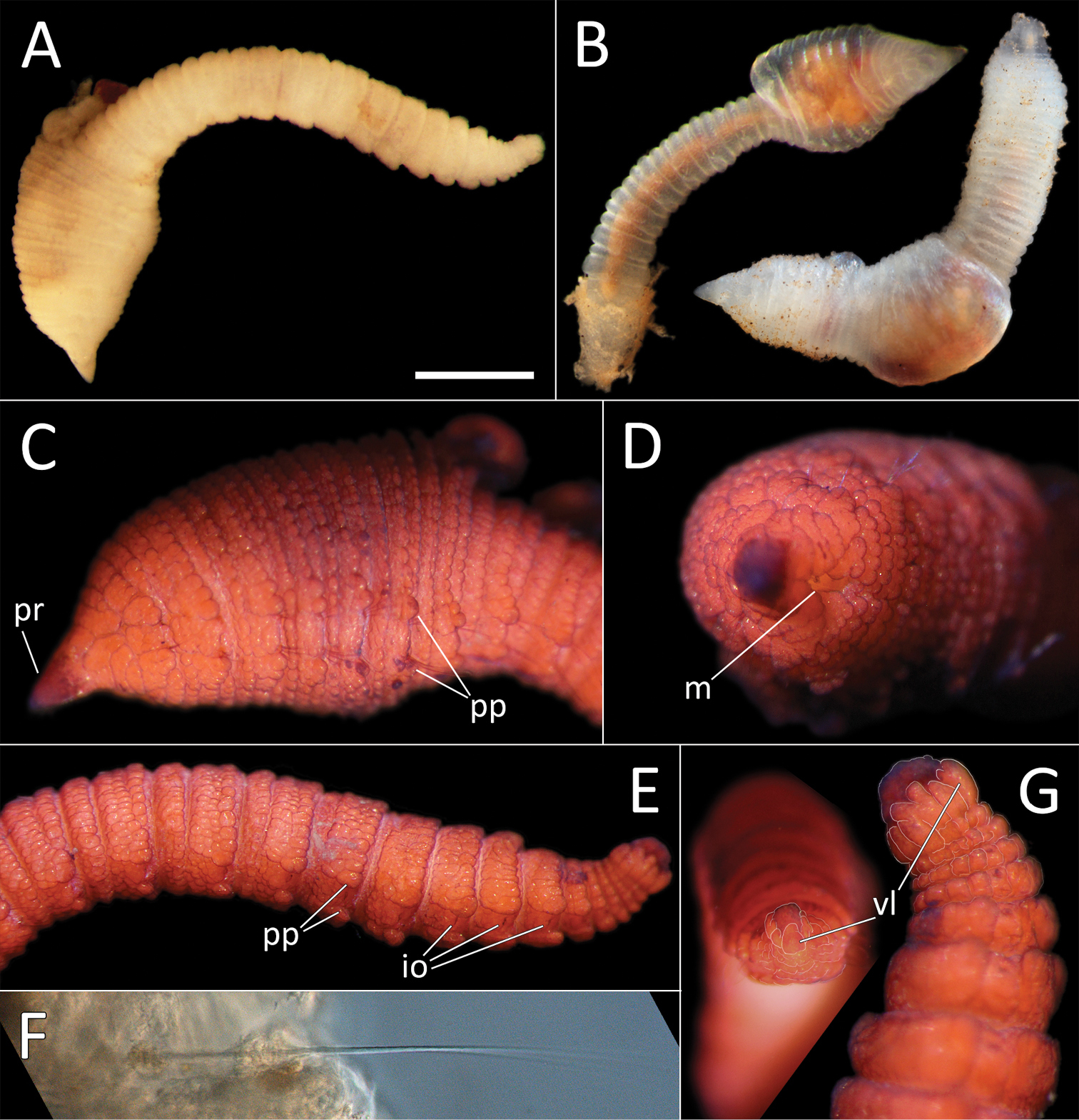
Travisia zieglerae sp. nov. A Lab image, whole specimen, pre-stain (holotype [specimen NHM_1431]) B Live images, whole specimens (specimen NHM_1911 [left], specimen NHM_188 [right]) C Lab image, lateral anterior, (holotype, stained, pr = prostomium, pp = parapodial lappets) D Lab image, distal anterior, (holotype, stained, m = mouth) E Lab image, lateral posterior, (holotype, stained, pp = parapodia, io = interramal organs) F Detail of capillary chaeta (paratype NHM_140) G Lab image, pygidium, distal view (left) and lateral view (right), with pygidial features outlined in a fine white line (holotype, stained, vl = ventral lobe). Scale bar: 1 mm (A). |