|
||
|
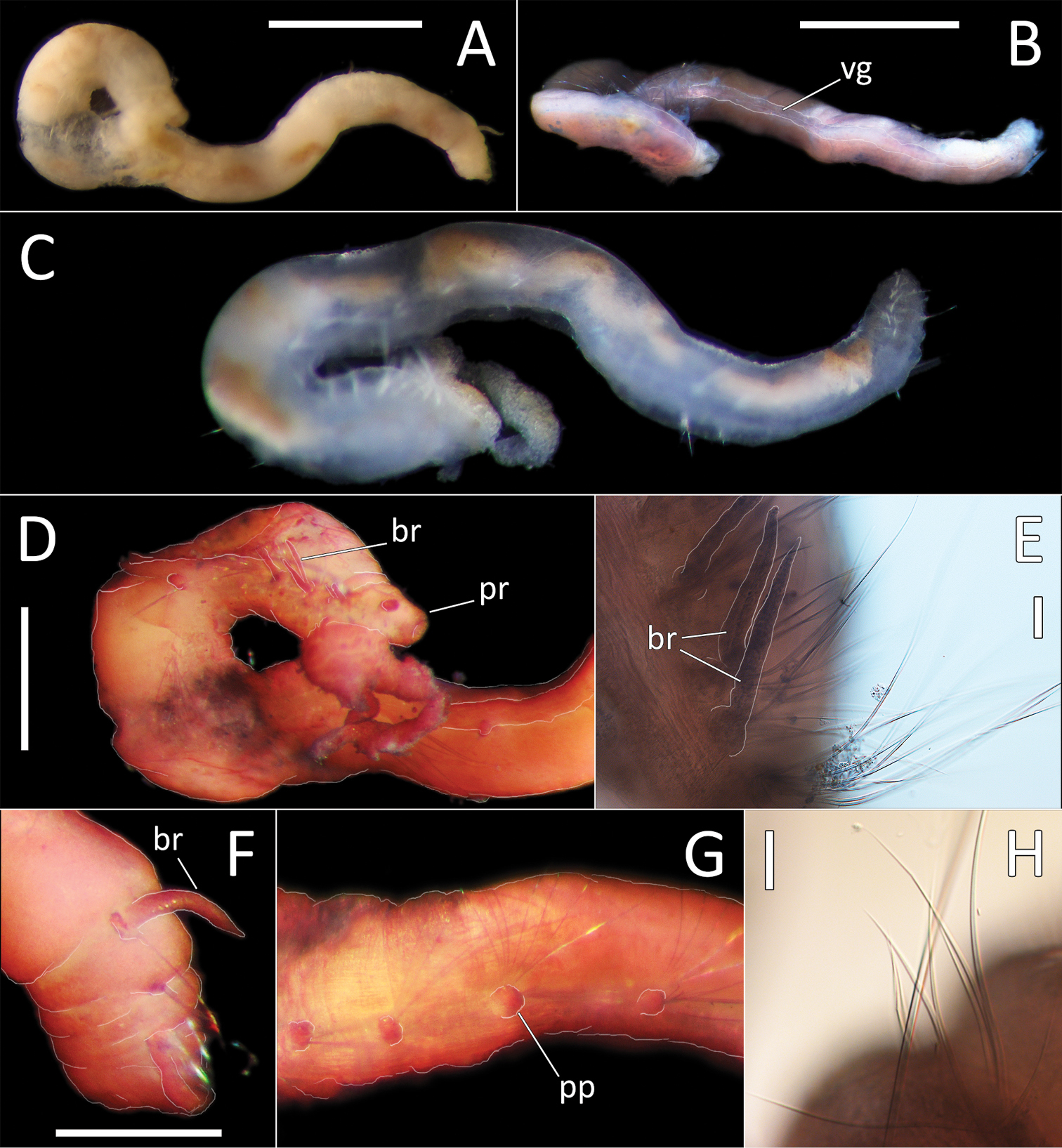
Ophelina sp. NHM_689 (specimen NHM_689). A Lab image, whole specimen, lateral view (pre-stain) B Lab image, whole specimen, ventral view (faded stain, vg = ventral groove) C Live image, whole specimen D Lab image, damaged anterior (stained, br = branchiae, pr = prostomium) E Lab image, detail of anterior branchiae (br = branchiae) F Lab image, posterior branchiae (stained, br = branchiae) G Lab image, mid-body parapodia (stained, pp = parapodia) H Lab image, detail of capillary chaetae. Morphological features in plates B, D–G have been outlined with a fine white line to improve clarity of those features. Scale bars: 1 mm (A, B); 0.5 mm (D); 50 μm (E); 0.25 (F, H). |