|
||
|
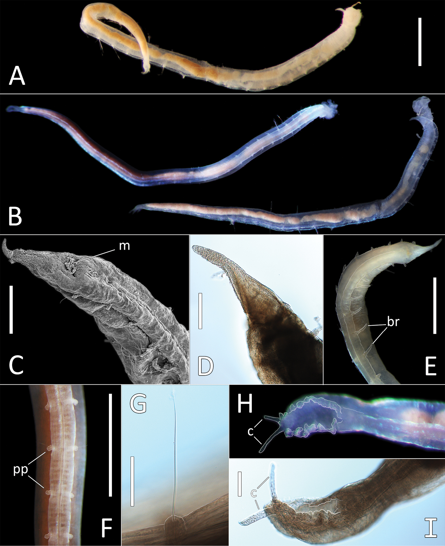
Ophelina nunnallyi sp. nov. A Lab image, whole specimen (paratype NHM_700) B Live images, whole specimens (holotype [specimen NHM_683] [left], paratype NHM_700 [right]) C SEM image, anterior and palpode (specimen NHM_1309A, m = mouth) D Lab image, detail of palpode, (paratype NHM_1273) E Lab image, anterior and branchiae (paratype NHM_1273, br = branchiae) F Lab image, parapodia (holotype, pp = parapodia) G Lab image, capillary chaeta (holotype) H Live image, anal funnel (holotype, c = cirri) I Lab image, detail of anal funnel (holotype, c = cirri). Morphological features in plates E, F, H, I have been outlined with a fine white line to improve clarity of those features. Scale bars: 1 mm (A, E, F); 200 μm (C, D); 100 μm (G, I). |