|
||
|
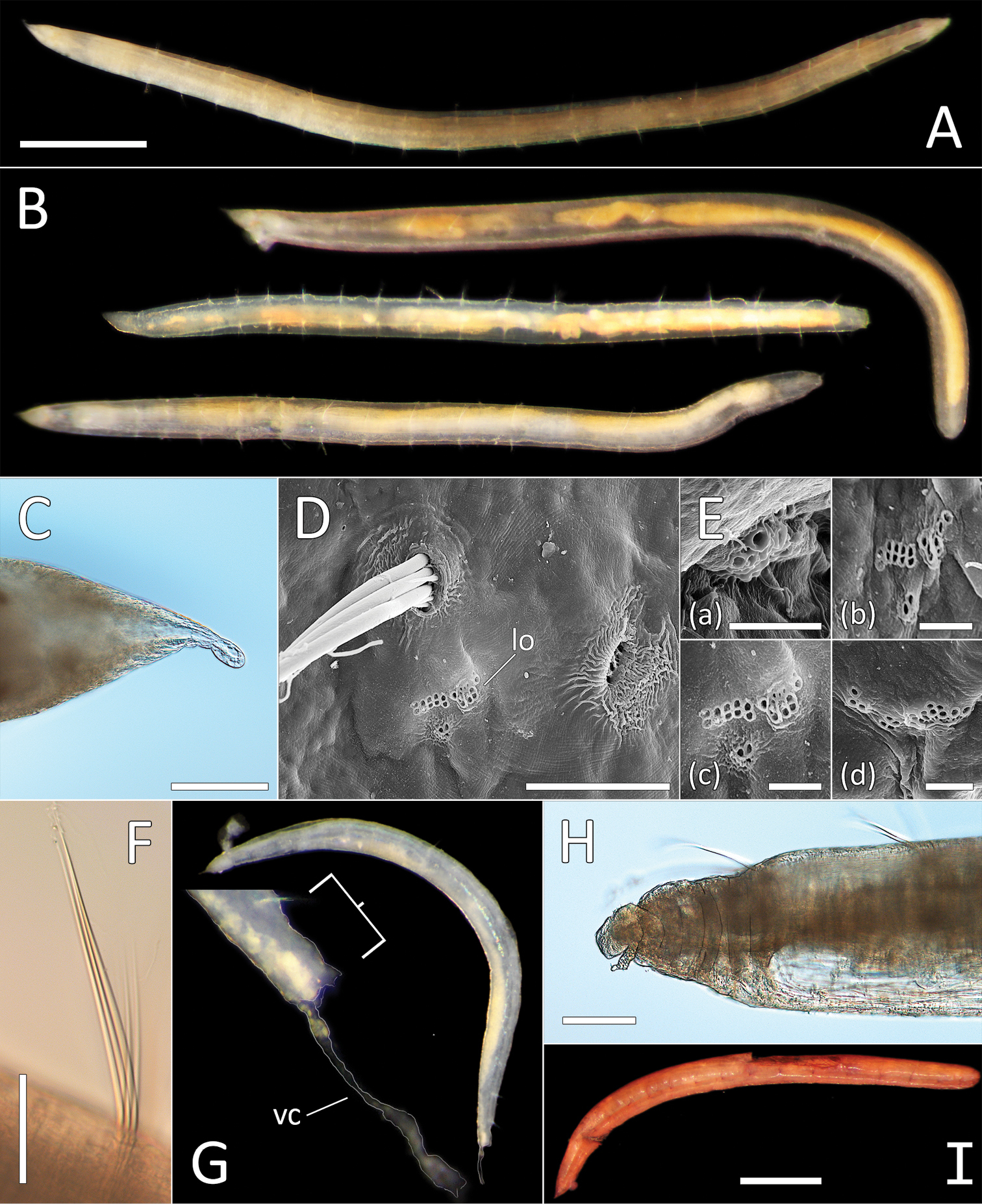
Ophelina ganae sp. nov. A Lab image, whole specimen (holotype [specimen NHM_1137] B Live images, whole specimens (holotype [bottom], paratype NHM_598 [middle], paratype NHM_1309 [top]) C Lab image, detail of palpode (holotype) D SEM image, second chaetiger with lateral organ (paratype NHM_1309, lo = lateral organ) E SEM images, lateral organs, (a) pre-chaetigerous segment, (b) chaetiger 1, (c) chaetiger 2, (d) chaetiger 17 (last chaetiger) (paratype NHM_1309) F Lab image, detail of capillary chaetae (holotype) G Live image, with detail of potential anal funnel (specimen NHM_473, vc = potential ventral cirrus). H Lab image, detail of posterior (holotype) I Lab image, whole specimen (paratype NHM_598, stained). Morphological features in plates G, H have been outlined with a fine white or black line to improve clarity of those features. Scale bars: 1 mm (A); 100 μm (C); 20 μm (D); 5 μm (E); 50 μm (F); 100 μm (H); 1 mm (I). |