|
||
|
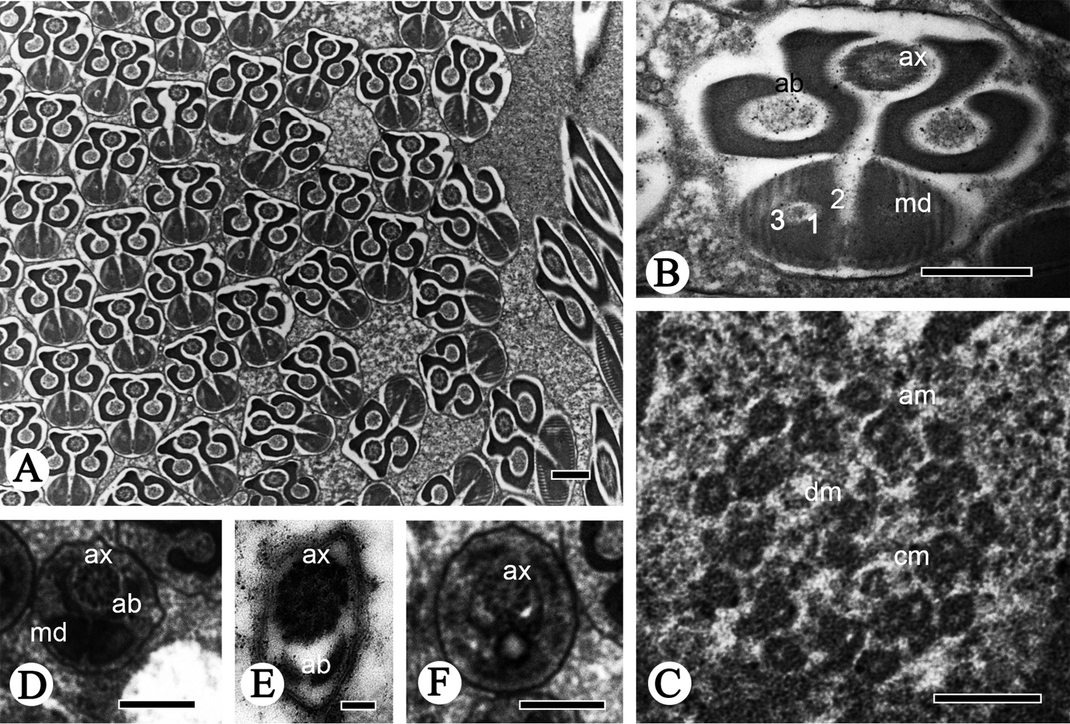
Cross-sections of the sperm flagellum of R. speculum. A, B Flagella, showing the axoneme (ax), accessory bodies (ab) and mitochondrial derivatives (md) including oval electron-lucid portion (1), an electron-dense region (2), and one mitochondrial cristae region (3) C axoneme, showing the typical 9 + 9 + 2 pattern, nine outermost accessory microtubules (am), a pair of central microtubules (cm), and doublet microtubules (dm) in between D flagellum, showing the axoneme (ax), accessory bodies (ab) and mitochondrial derivatives (md) E–F flagellum without mitochondrial derivatives (md). Scale bars: 0.5 µm (A, B, D, F); 0.1 µm (C, E). |