|
||
|
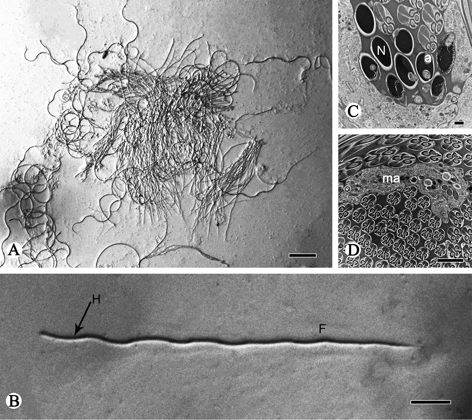
TEM and light micrographs of spermatozoon and spermatodesms of R. speculum. A Light micrograph of spermatodesm B light micrograph of spermatozoon with the head (H, arrow) and flagellum (F) C, D cross-sections of the oval nucleus (N), showing the acrosome (a) and homogenous matrix (ma). Scale bars: 50 µm (A); 20 µm (B); 0.5 µm (C); 2 µm (D). |