|
||
|
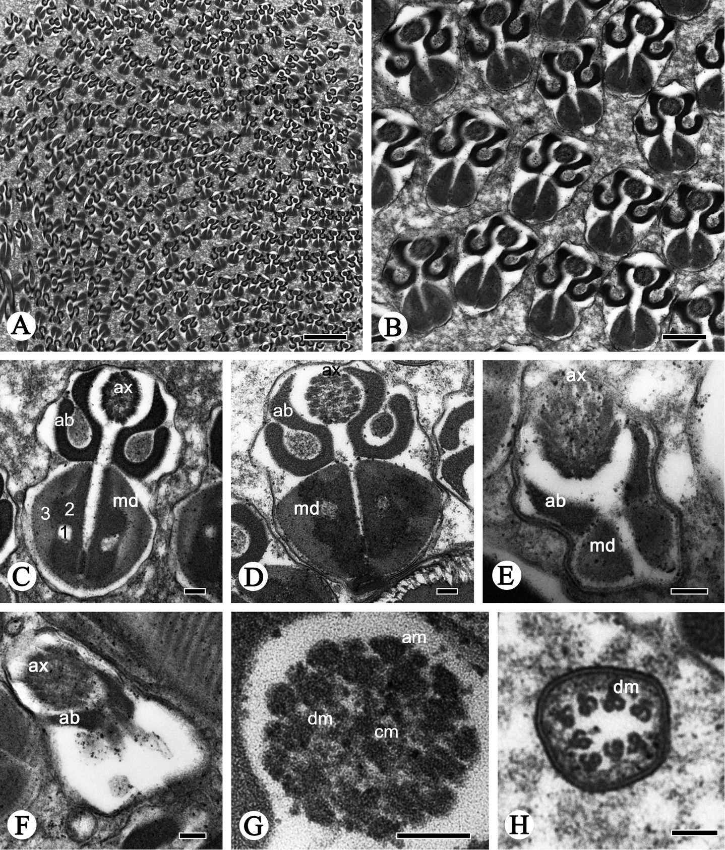
Cross-sections of the sperm flagellum of P. shantungensis. A–D Flagella, showing axoneme (ax), fishhook-shaped accessory bodies (ab), D-shaped mitochondrial derivatives (md), containing oval lucent region (1), serrated electron-dense region (2) and mitochondrial cristae region (3) E–F flagellum, mitochondrial derivatives slowly disappear, axonemes (ax) become disordered, accessory bodies (ab) become smaller G axoneme, showing the typical 9 + 9 + 2 pattern, nine outermost accessory microtubules (am), nine doublet microtubules (dm) and two innermost central microtubules (cm) H Showing doublet microtubules finally disappearing. Scale bars: 2 µm (A); 0.5 µm (B); 0.1 µm (C–H). |