|
||
|
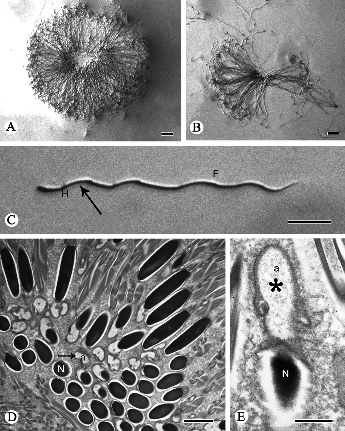
TEM and light micrographs of spermatozoa and spermatodesms of P. shantungensis. A, B Light micrographs of spermatodesm and spermatozoa C light micrograph of a single spermatozoon with the head (H, arrow) and wavy flagellum (F) D, E TEM micrographs of cross-sections of spermatozoa, showing the acrosome (a) and nucleus (N). Arrow shows head cluster, asterisk indicates the acrosome. Scale bars: 20 µm (A–C); 2 µm (D); 0.5µm (E). |