|
||
|
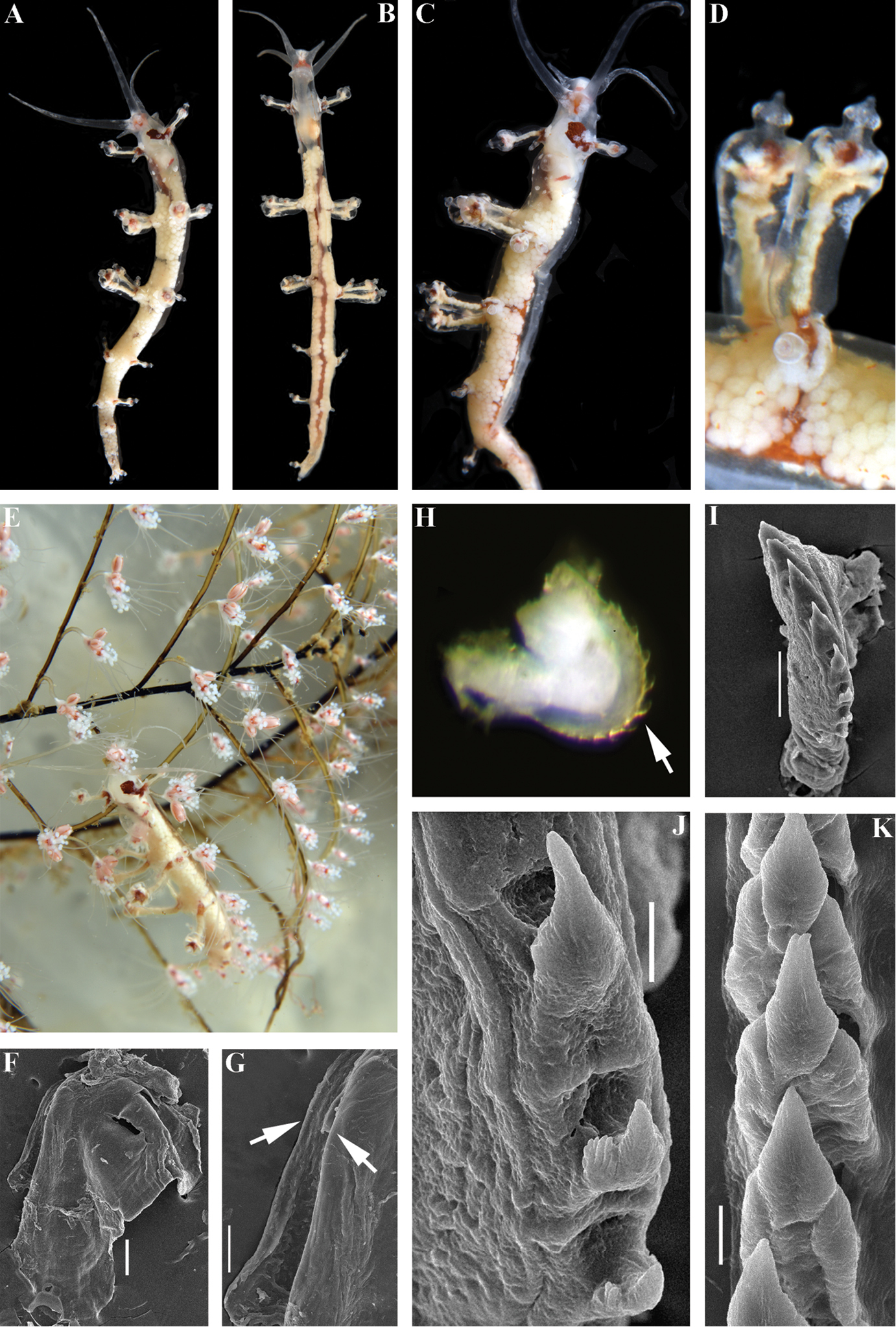
Myja hyotan sp. n., holotype. A dorsal view B ventral view C lateral view D details of cerata E dorsal view on hydroids in situ F jaw G smooth masticatory processes of jaws (indicated by arrows), SEM H radula on odontophore, to show reduced anteriormost teeth (arrow), LM I anterior teeth with strongly reduced anteriormost teeth, SEM J teeth from the middle part of radula K posterior part of radula to show smooth teeth. Scale bars: 100 μm (F); 50 μm (G, I); 10 μm (J, K). Photographs of living specimens by TA Korshunova and AV Martynov, SEM images by AV Martynov. |