|
||
|
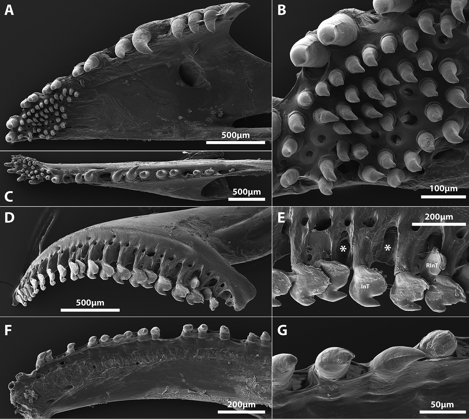
Scanning electron micrographs of the tooth-bearing oral jaw bones of Aspasmogaster costata (AMS I.19103-015, 32.0 mm SL) and Lepadichthys coccinotaenia (SAIAB 49396, 31.0 mm SL). A Premaxilla of A. costata, right side in ventral view (image reversed) B Close up of lingual toothpatch on premaxilla of A. costata shown in A C Dentary of A. costata, right side in ventral view (image reversed) D Premaxilla of Lepadichthys coccinotaenia, right side in lateral view (image reversed) E Close up of incisiviform teeth located on posterior part of premaxilla of L. coccinotaenia shown in D Asterisks (*) highlight locations of crypts associated with dislodged replacement teeth F Dentary of Lepadichthys coccinotaenia, right side in medial view G Close up of conical teeth located along midregion of dentary of L. coccinotaenia shown in F Abbreviations: InT, incisiviform tooth; RInT, replacement incisiviform tooth. |