|
||
|
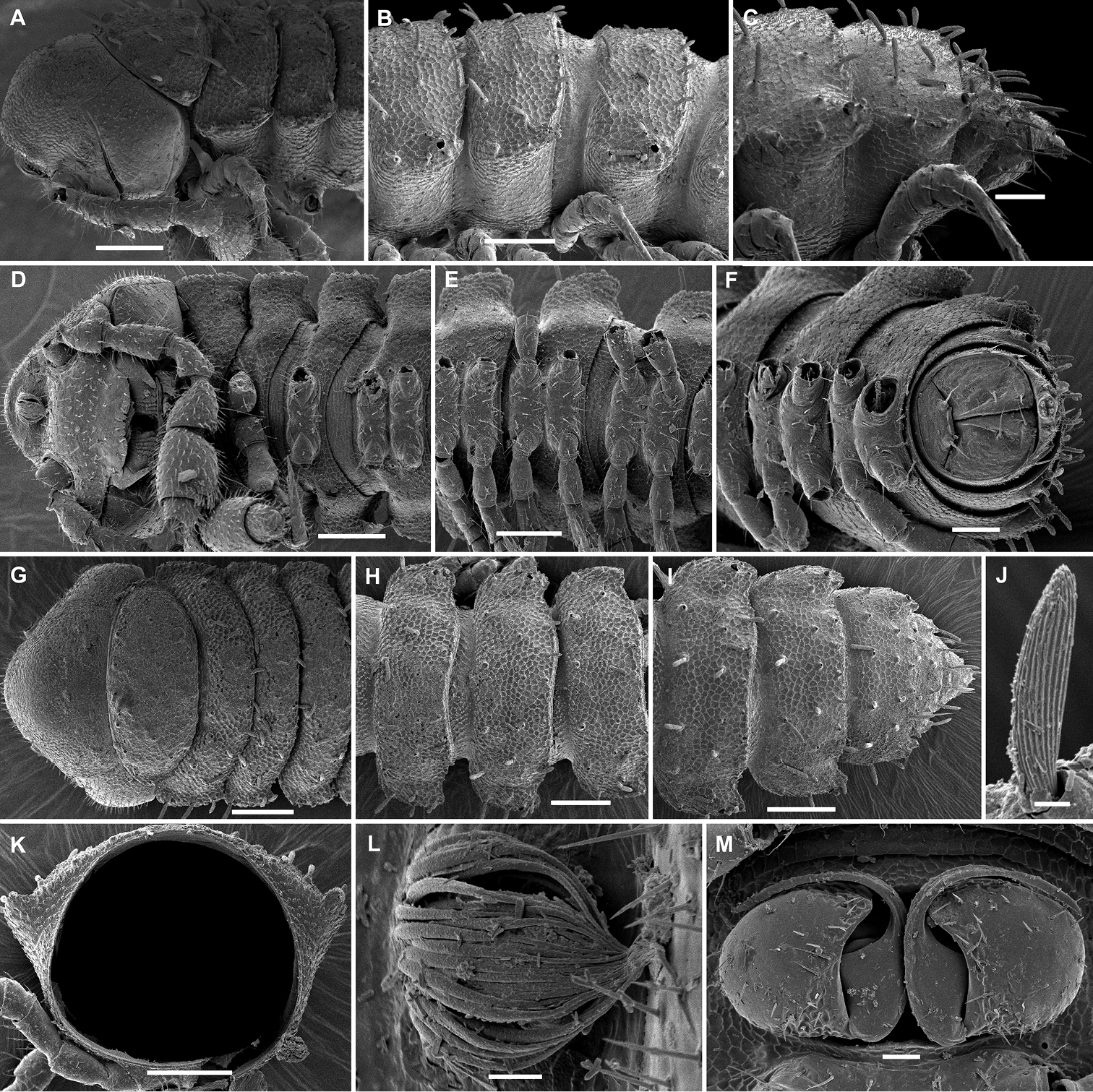
Hemisphaeroparia falcata sp. n., SEM micrographs of ♂ paratype A, D, G anterior part of body, lateral, ventral and dorsal views, respectively B, E, H midbody segments, lateral, ventral and dorsal views, respectively C, F, I posterior part of body, lateral, ventral and dorsal views, respectively J tergal seta, lateral view K midbody segment, caudal view L epicranial tubercle with filaments M both gonopods in situ, ventral view. Scale bars: 0.1 mm (A, B, D, E, G–I, K), 0.05 mm (C, F), 0.02 mm (M), 0.01 mm (L), 0.005 mm (J). |