|
||
|
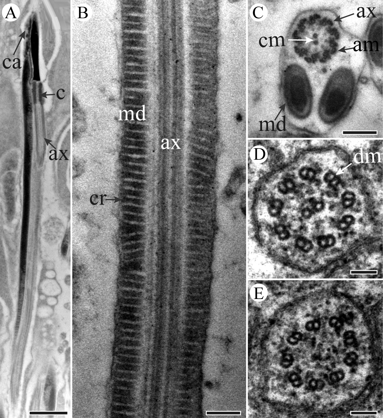
TEM micrographs of sperm tail region of P. kaempferi. A Longitudinal section through the neck and tail regions, showing nucleus (n), centriolar adjunct (ca), mitochondrial derivative (md), centriole (c) and axoneme (ax) B Higher magnification of longitudinal section of sperm tail, showing axoneme (ax) and mitochondrial derivatives with cristae (cr) C Cross-section of tail region, showing two mitochondrial derivatives (md) and a 9 + 9 + 2 microtubular pattern (i.e., 9 accessory microtubules (am), 9 double microtubules (dm), and two central microtubules (cm)) axoneme (ax) D Higher magnification of cross-section of axoneme (ax), showing 9 double microtubules (dm), and two central microtubules left E Higher magnification of cross-section of axoneme (ax) showing only 9 double microtubules (dm) remained. Scale bars: 1 μm (A), 200 nm (B, C), 100 nm (D, E). |