|
||
|
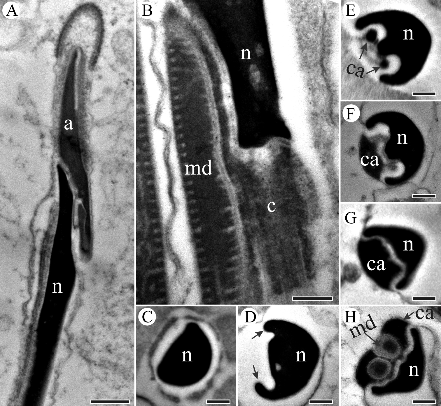
TEM micrographs of sperm neck region of P. kaempferi. A Longitudinal section showing head region, showing conical acrosome (a) and tapered nucleus (n) B Longitudinal section of nucleus-flagellum transition region, showing nucleus (n), mitochondrial derivative (md) and centriole (c) C Cross-section of nucleus (n) with a deltoid appearance D Cross-section through the mid-neck region, showing an invagination at one side of the nucleus (n) developing two ridges (arrowed) E–G Cross-sections through neck region, showing centriolar adjunct (ca) and nucleus (n). H Cross-section through the mid-neck region, showing nucleus (n), mitochondrial derivatives (md) and centriolar adjunct (ca). Scale bars: 500 nm (A), 200 nm (B–H). |