|
||
|
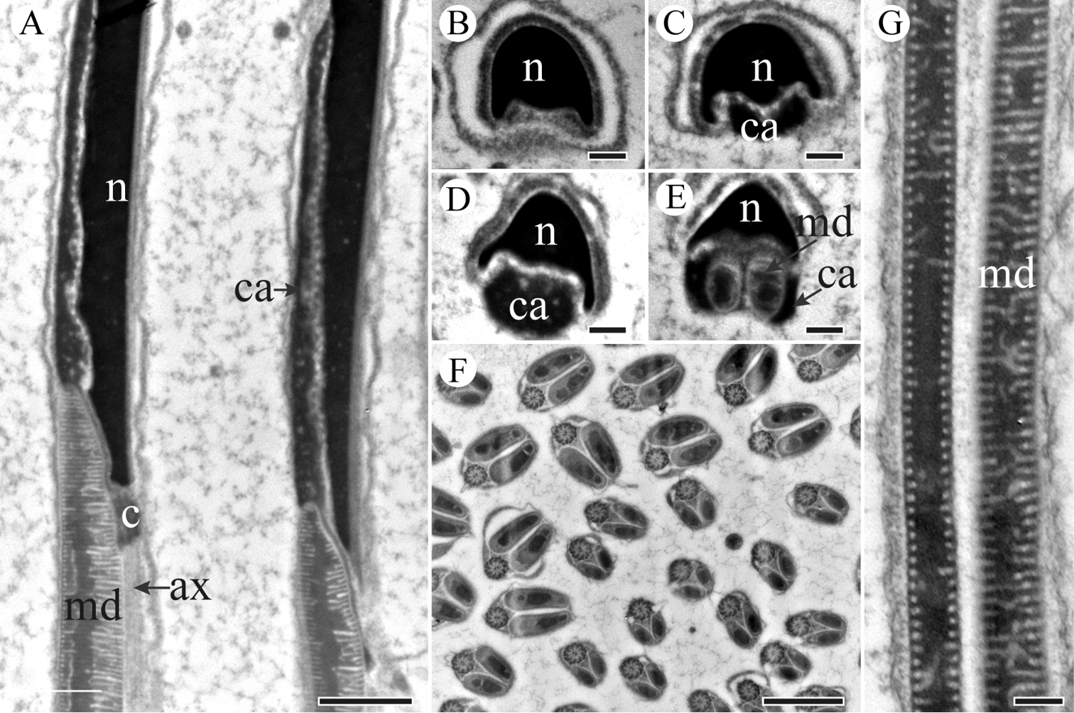
TEM sections through the neck and tail regions of the spermatozoa of K. caelatata. A Longitudinal section through the neck region showing nucleus (n), centriolar adjunct (ca), centriole (c), axoneme (ax) and mitochondrial derivatives (md) B Cross-section through the mid-neck region, showing one side of the nucleus forms two ridges. C and D Cross-sections through the posterior part of nucleus, showing centriolar adjunct (ca) flanked nucleus (n) E Cross-section of the base of the nucleus, showing triangular nucleus (n) and two mitochondrial derivatives (md) embedded into the material of the centriolar adjuncts (ca) F Cross-section through sperm tails, showing mitochondrial derivatives with distinct diameters G Longitudinal section of sperm tail, showing paired mitochondrial derivatives (md). Scale bars: 1 μm (A, F), 200 nm (B–E, G). |