|
||
|
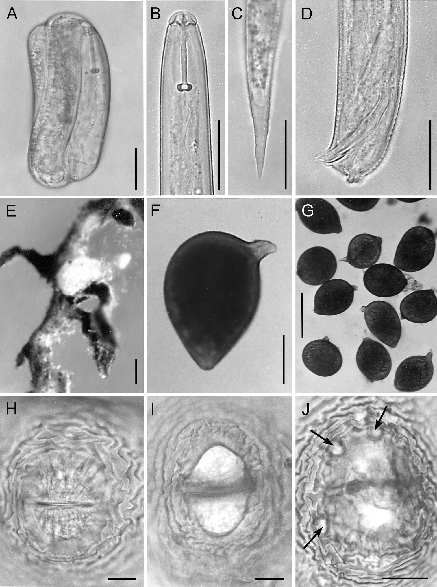
LM micrographs of Heterodera fici from Italy. A Embryonated egg with evident second stage juvenile stylet B Second stage juvenile anterior end C Second stage juvenile tail D Male tail with the characteristic tail tip E Females on Ficus carica roots F whole body of newly formed cyst G Females and cysts H-J Vulval cone structures, with clear illustration of vulval slit (in H), fenestral area (in I) and bullae (in J). Scale bars: 20 µm (A-D, H-J); 200 µm (F); 500 µm (E, G). |