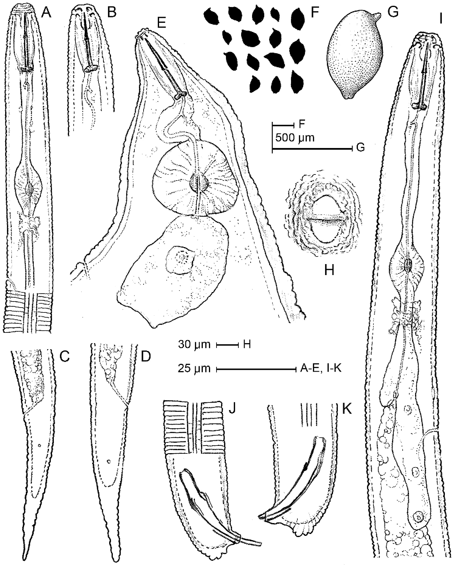
Line drawings of Heterodera fici from Italy. A, B Anterior body portions of second stage juvenile C, D Second stage juvenile tail E Female anterior region F, G Cyst shape H Fenestral structures J, K Male tail, showing spicules and cloacal tube I Male pharyngeal region.


  Part of: Fanelli E, Vovlas A, Santoro S, Troccoli A, Lucarelli G, Trisciuzzi N, De Luca F (2019) Integrative diagnosis, biological observations, and histopathology of the fig cyst nematode Heterodera fici Kirjanova (1954) associated with Ficus carica L. in southern Italy. ZooKeys 824: 1-19. https://doi.org/10.3897/zookeys.824.26820