|
||
|
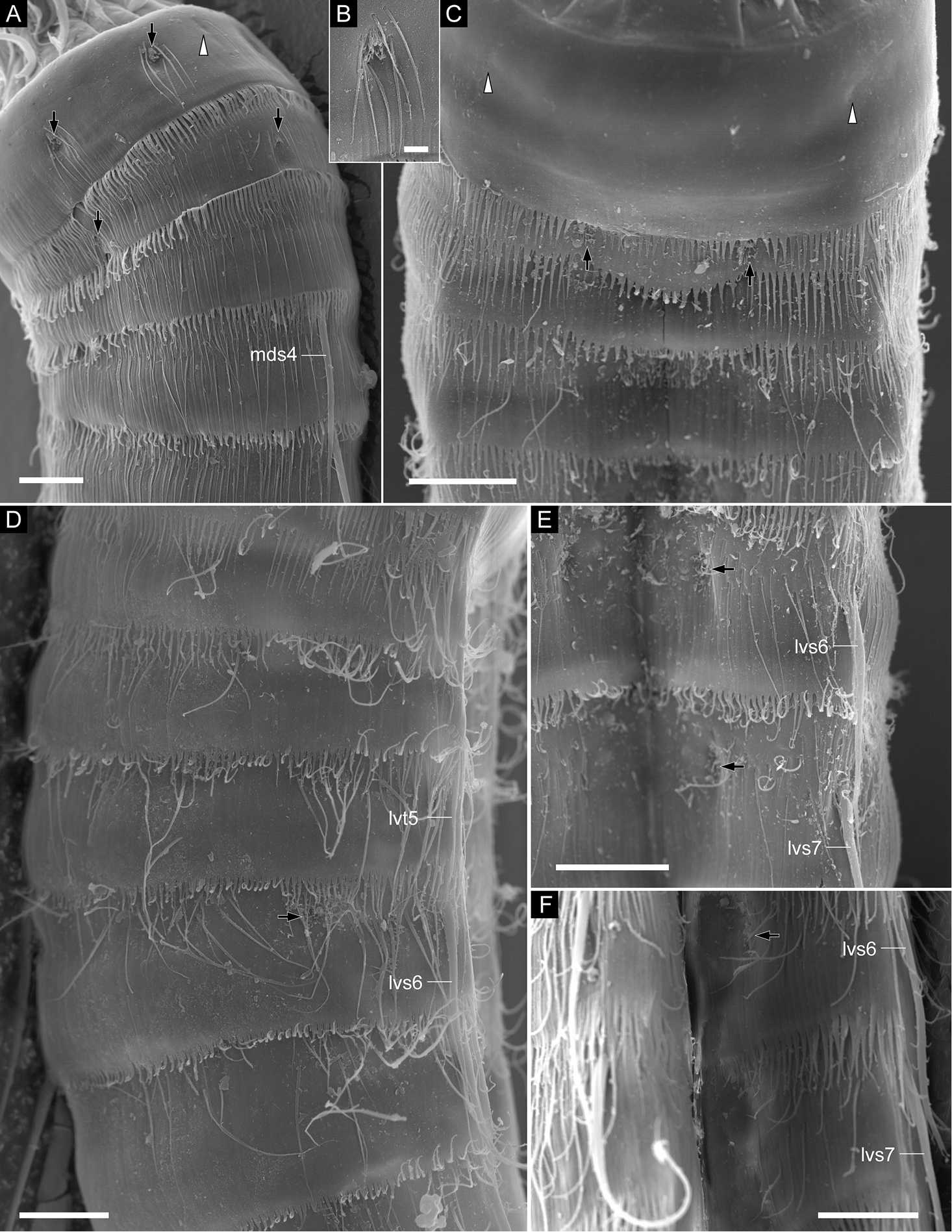
Echinoderes pterus sp. n., scanning electron micrographs. Females (A, BZMB 11669a, collected at station 152-1 (Karasik Seamount) DZMB 11661c, collected at station 66 (Mediterranean deep sea of Crete) FZMB 11669b, collected at station 152-1 (Karasik Seamount)) and a male (C, EZMB 11661a, collected at station 66 (Mediterranean deep sea off Crete)). A segments 1–4, laterodorsal view (left side) B close-up of laterodorsal sensory spot on segment 1 C segments 1–4, ventral view D segments 3–7, lateral view (right side) E sternal plates on segments 6 and 7 F sternal plates on segments 6 and 7. Abbreviations: lvs, lateroventral acicular spine; lvt, lateroventral tube; mds, middorsal acicular spine. Digits after abbreviations indicate the corresponding segment number. Black arrows point to sensory spots; white arrowheads mark type-1 gland cell outlets. Scale bars: 10 µm (A, C–F), 2 µm (B). |