|
||
|
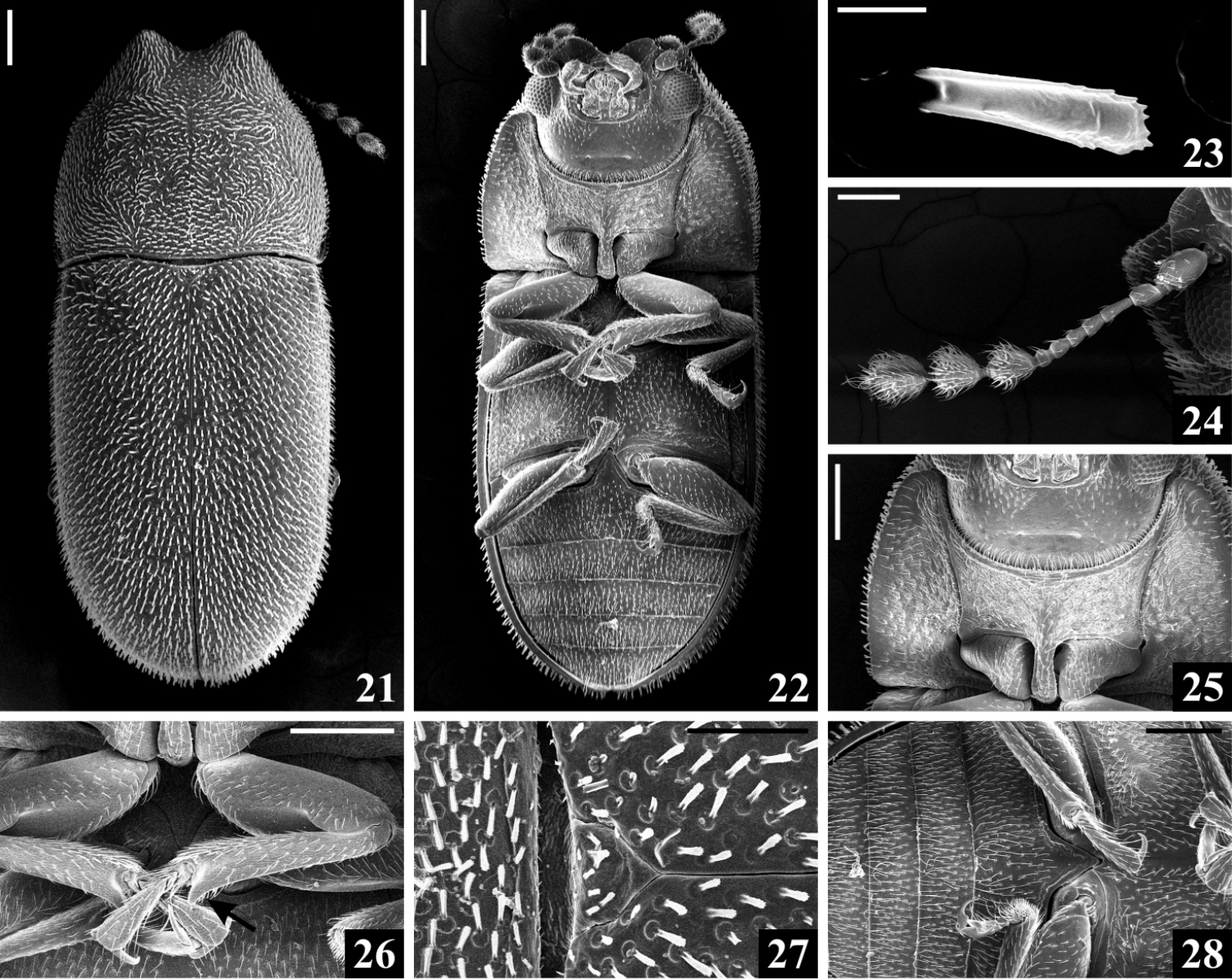
Cis pallidus Mellié, 1849, scanning electron microscopy. 21 Dorsal view 22 Ventral view 23 Elytral bristle 24 Antenna 25 Part of head and prothorax in ventral view 26 Protibiae, showing the outer apical angle (arrow) 27 Part of pronotum and elytra, with scutellar shield 28 Part of metaventrite and abdominal ventrites. Scale bars: 0.2 mm (21–22, 25–26, 28), 0.01 mm (23), 0.1 mm (24, 27). |