|
||
|
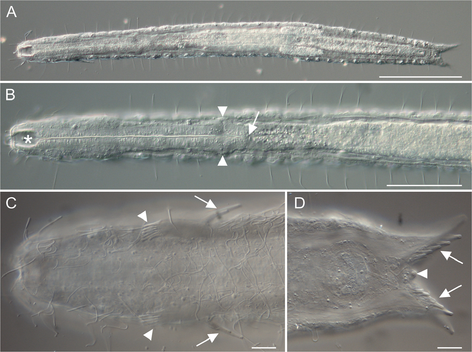
Paraturbanella xaymacana sp. n., holotype. Differential interference contrast photomicrographs. A Habitus, ventral view B Anterior region, ventral view, showing the buccal cavity (asterisk), the pharyngeal pores (arrowheads), and the pharyngo-intestinal junction (arrow) C Anterior region, ventral view, showing the lateral and ventral ciliation, the anterior adhesive tubes (arrowheads), and the additional adhesive tubes (Seitenfüsschen) (arrows) D Posterior region, ventral view, showing the medial cone (arrowhead) and the posterior adhesive tubes (arrows). Scale bars: 100 μm (A), 50 μm (B), 20 μm (C–D). |