|
||
|
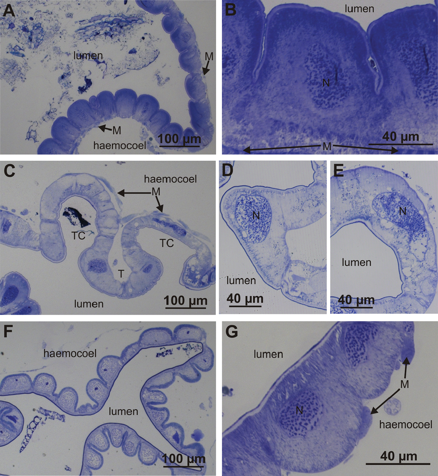
Histological structure of the hindgut epithelium in the anterior chamber and papillate region. A Ventral-lateral epithelium in the anterior chamber B Apical parts of ventral and lateral epithelial cells are bulging into the hindgut lumen C Dorsal epithelium in the anterior chamber forms typhlosole (T) and two typhlosole channels (TC) D Epithelial cell of typhlosole E Epithelial cell of typhlosole channel F Hindgut epithelium in the papillate region G Basal parts of epithelial cells in the papillate region are bulging into haemocoel. Abbreviations: M – muscles, N – cell nucleus. |