|
||
|
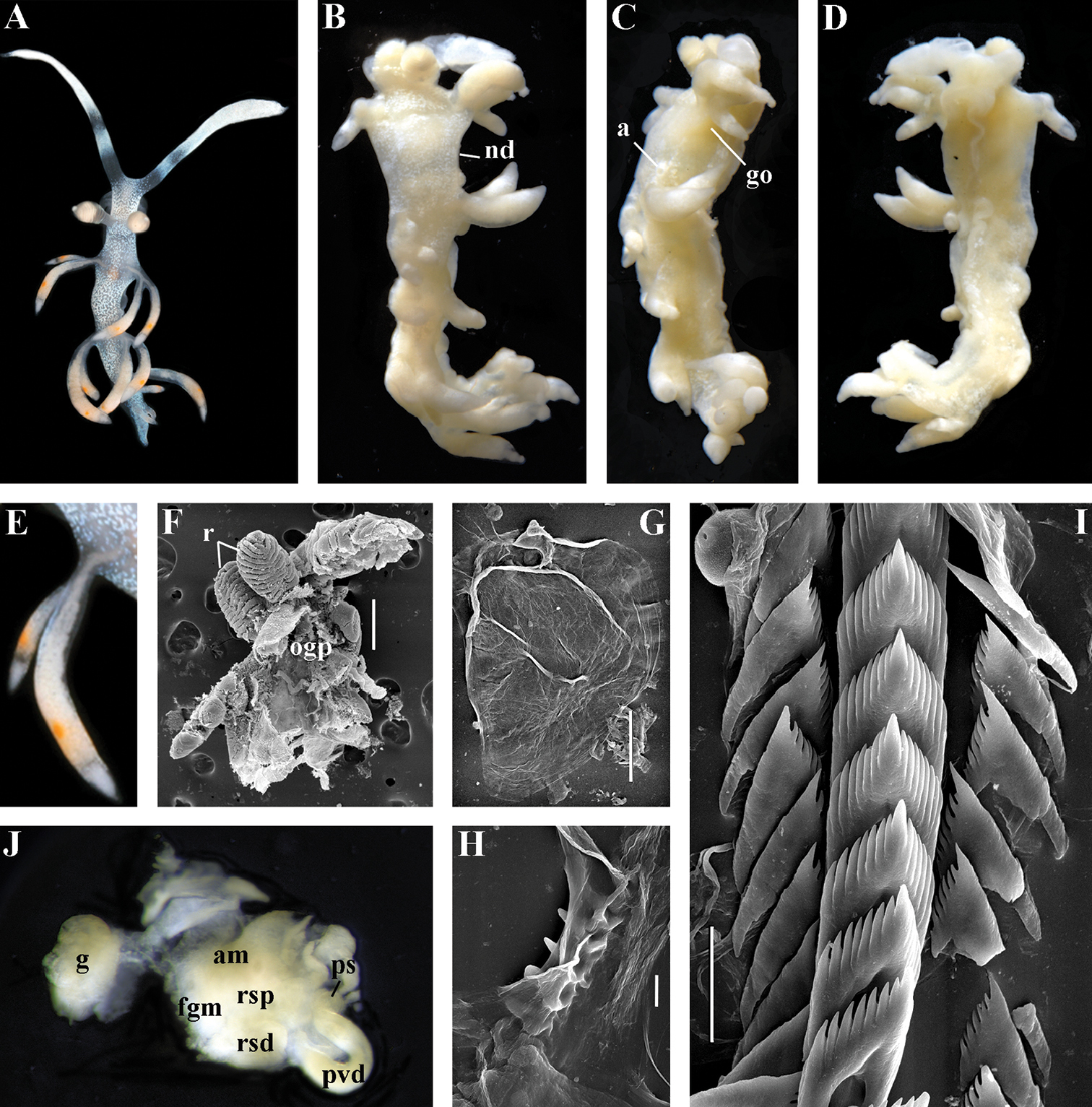
Samla bicolor (Kelaart, 1858). Vietnam, Nhatrang Bay. ZMMU Op-68, living specimen 5 mm in length: A dorsal view B dorsal view (fixed) C lateral view (fixed) D ventral view (fixed) E details of cerata F dissected anterior part (pharynx removed) and rhinophores, SEM G jaw, SEM H details of masticatory process of jaw, SEM I radular teeth, posterior part, SEM J reproductive system, light microscopy. Abbreviations: a anus am ampulla fgm female gland mass g gonad go genital opening nd discontinuous notal edge ogp oral gland penetrating into basis of cerata ps penial sheath pvd prostatic vas deferens r rhinophores rsd distal receptaculum seminis rsp proximal receptaculum seminis. Scale bars: F = 300 μm; G = 100 μm; H = 10 μm; I = 30 μm. Photos of living specimens by O.V. Savinkin, other photos and SEM images by A.V. Martynov. |