|
||
|
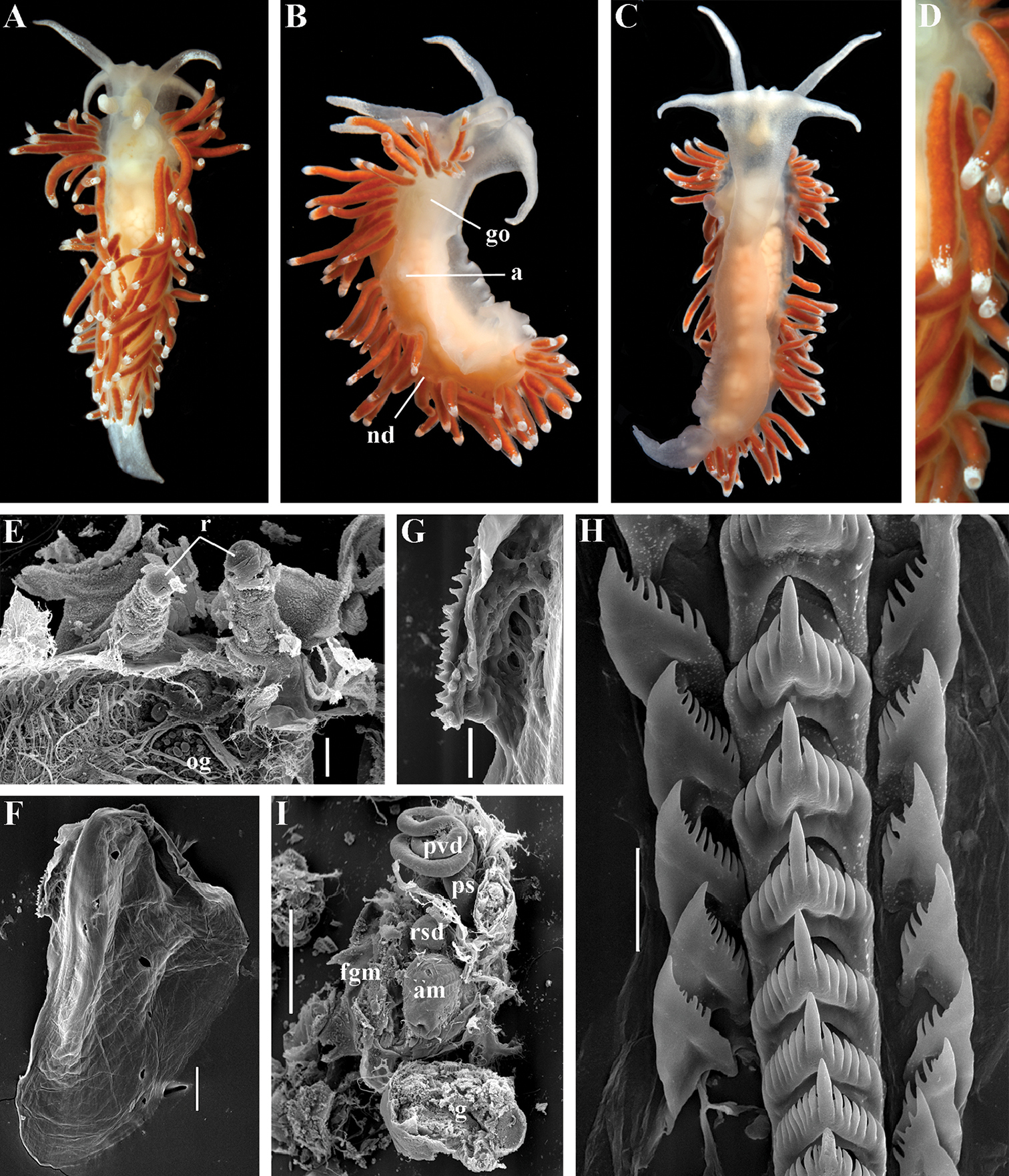
Microchlamylla gracilis gracilis (Alder & Hancock, 1844), comb. n. Norwegian Sea, Gulen Dive Center. ZMMU Op-502, living specimen 23 mm length: A dorsal view B lateral view C ventral view D details of cerata E dissected anterior part (pharynx removed) and rhinophores, SEM F jaw, SEM G details of masticatory process of jaw, SEM H radular teeth, posterior part, SEM I reproductive system, SEM. Abbreviations: a anus am ampulla fgm female gland mass go genital opening nd discontinuous notal edge og oral glands ps penial sheath pvd prostatic vas deferens r rhinophores rsd distal receptaculum seminis. Scale bars: E, I = 1 mm; F = 300 μm; G, H = 30 μm. Photos and SEM images by T.A. Korshunova, A.V. Martynov. |