|
||
|
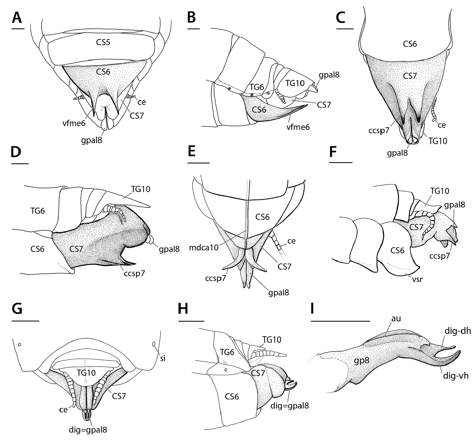
Annotated illustrations of digging structures on female genitalia. A–BEremiaphilidae-type digging spine: Eremiaphilasp. terminalia A ventral view B lateral view. C–D Rivetina-type digging spine: Rivetina Berland & Chopard, 1922 terminalia C ventral view D lateral view. E–F Chroicoptera-type digging spine: Chroicoptera longa Giglio-Tos, 1915 terminalia (E) ventral view; Chroicoptera saussurei Giglio-Tos, 1915 F lateral view. G–I Ligaria-type digging spine: Ligaria Stål, 1877 G dorsal view H lateral view I lateral view (left side) of the left gonapophysis 8. Note that in the Ligaria-type, the digging structures are gonapophyses 8. Figures A–E and G–I reproduced and adapted from Wieland (2013); figure F reproduced from Wieland 2013, which was modified from Kaltenbach 1996. Abbreviations: au = aulax; ccsp7 = centrocoxosternal processes 7; ce = cercus; CS5–7 = coxosternite 5–7; dig-dh = digging device, dorsal hook; dig-vh = digging device, ventral hook; mdca10 = middorsal carina 10; vfme6 = ventromedian expansion (of ventral fold); gpal8 = apical lobe of gonapophysis 8; si = spiracle; TG6–10 = tergite 6–10. Scale bars: 1 mm. |