|
||
|
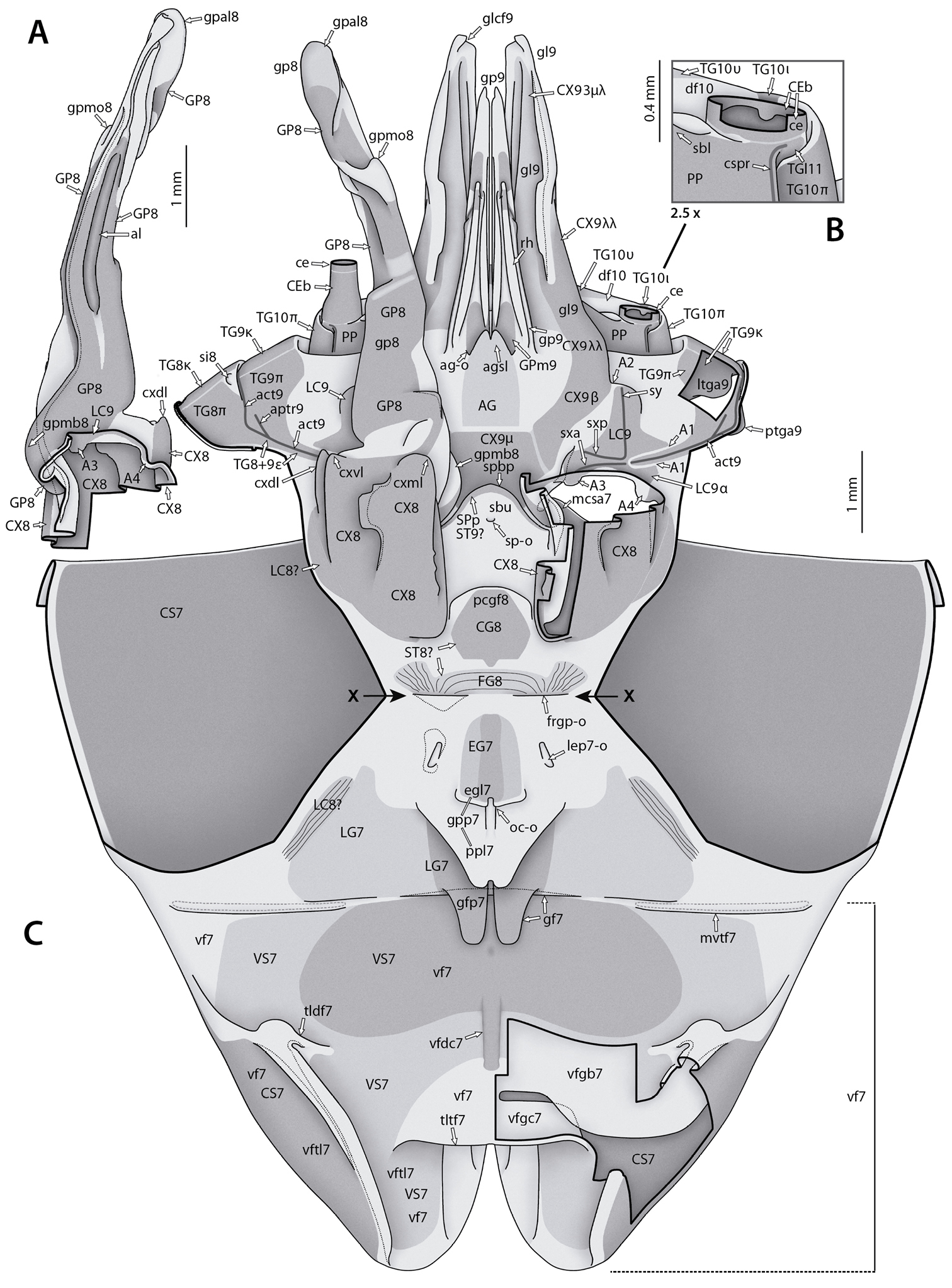
Annotated illustrations of the female Sphodromantissp. postabdomen and genitalia for morphological use. A left gonapophysis 8 with adjoining parts of coxa 8 and laterocoxa 9 in dorsal view B lateral parts of terminal abdomen in ventral view, with focus on base of cercus (enlarged 2.5× from C) C entire genital region, most parts in external view. Posterior parts (segments VIII–XI) are bent 180° dorsally and to the anterior compared to anterior parts (of segment VII), along the axis marked by arrows X. The lower half of the illustration shows the ventral fold 7 (“subgenital lobe”) and the genital fold area in dorsal view; the upper half of the illustration shows the ventral sides of segments VIII–XI in ventral view. Left gonapophysis 8 removed. Thick black lines are (virtual) cutting lines. Continuous thin black lines are freely visible edges (= lines along which the cuticle bends away from the observer’s view). Dashed thin black lines are (parts of) edges hidden beneath other cuticle. Thick gray lines are internal ridges. Dashed gray line next to fold mvtf7 in C giving outline of area where dorsal and ventral walls of ventral fold 7 are firmly connected by columellae. Membranous cuticle in very light gray, sclerotized cuticle in medium to darker gray (darker = more strongly sclerotized); cuticle shaded darker where it goes underneath other cuticle. For abbreviations see text, glossary, or Suppl. material 2: Extended abdominal glossary. |