|
||
|
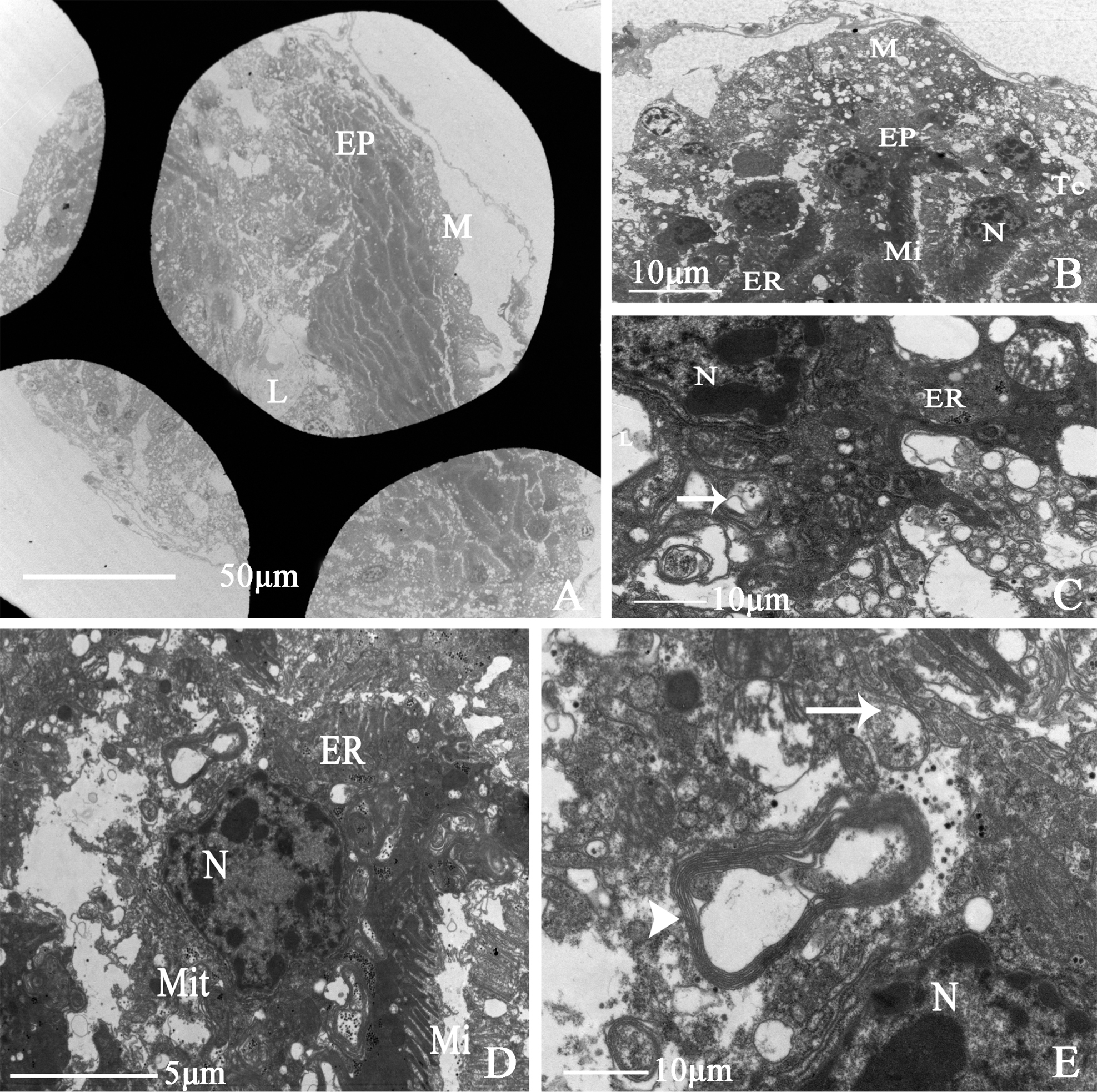
Ultrastructure of vagina of D. vulgaris (Dash & Viraktamath, 1998) A cross-section of vagina, showing (M) muscle sheath, (EP) epithelium, (L) lumen B, C showing (M) muscle sheath, (EP) epithelium, (ER) endoplasmic reticulum, (Mit) mitochondria, (Tc) tracheole, (Sj) the arrow indicates septate junction D showing (N) epithelial cell nucleus, (ER) endoplasmic reticulum, (Mit) mitochondria, (Mi) microvillus; (E) showing (N) epithelial cell nucleus, (Sj) the long arrow indicates the septate junction. |