|
||
|
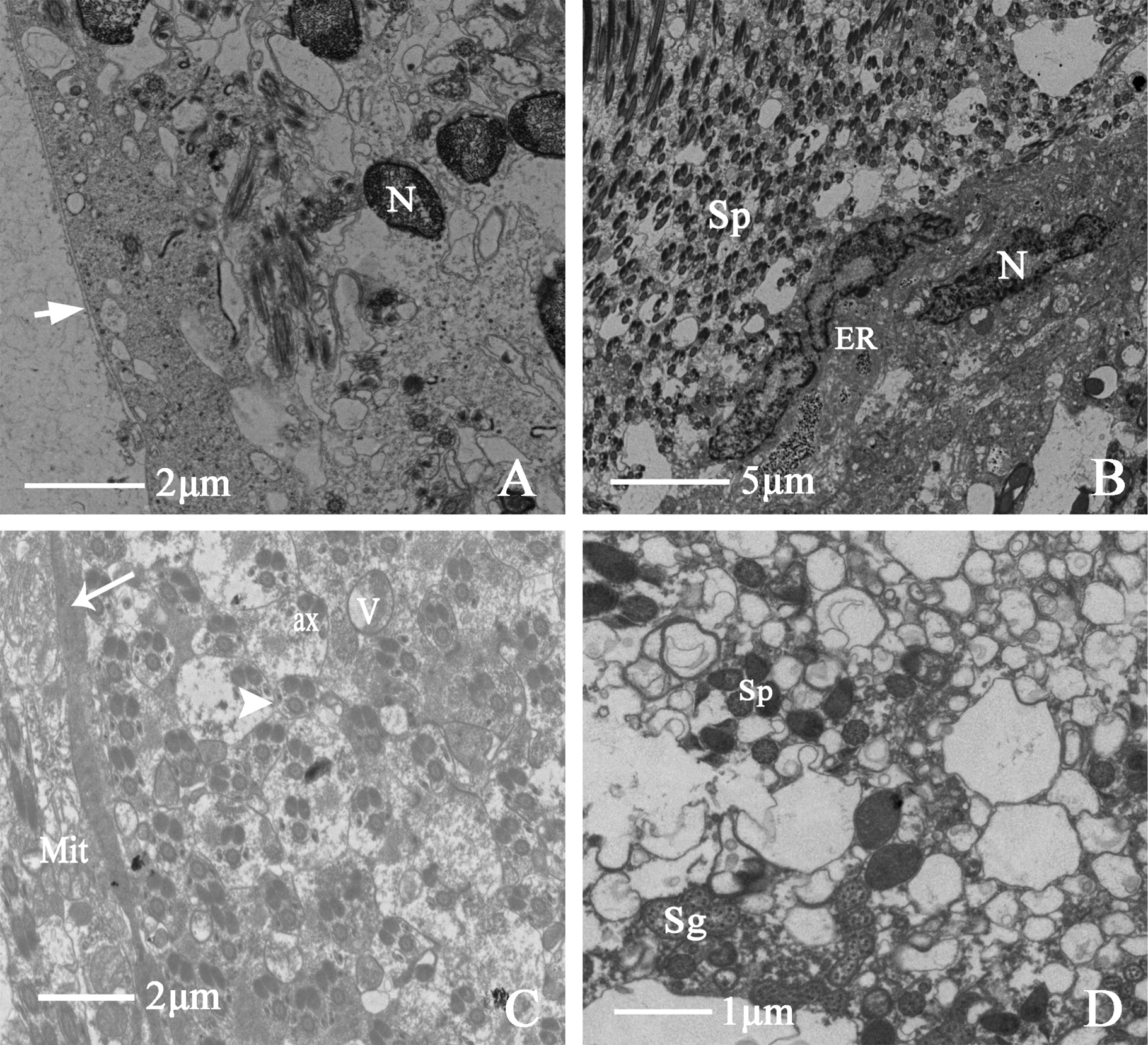
Ultrastructure of testicular follicle of D. vulgaris (Dash & Viraktamath, 1998) A cross-section of testicular follicle, showing (TM) testicular follicle membrane, (N) epithelial cell nucleus, (TM) the arrow indicates testicular follicle membrane B showing (Sp) spermatid, (ER) endoplasmic reticulum, (N) epithelial cell nucleus C showing (ax) axoneme, (V) vesicle, (Mit) mitochondria, (BM) the long arrow indicates thick basal membrane, (Sj) the triangular arrowhead indicates septate junction D showing (Sp) spermatid, (Sg) secretory granules. |