|
||
|
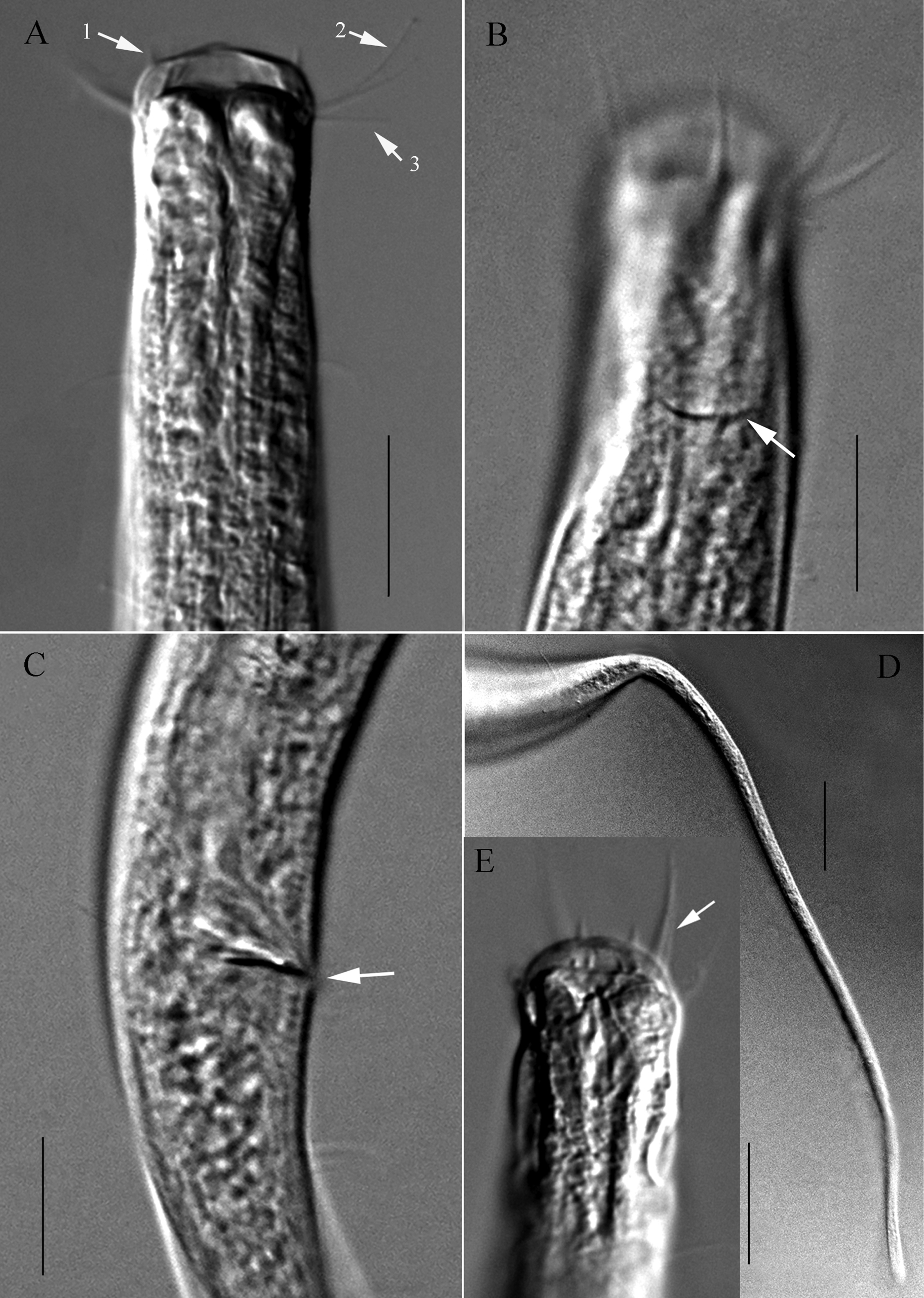
Microscopic images of Stylotheristus flagellicaudatus sp. nov. A lateral view of anterior portion of holotype, showing conical inner labial setae (arrow 1), outer labial setae (arrow 2), subcephalic setae (arrow 3) and buccal cavity B lateral view of anterior portion of holotype, showing cephalic setae and amphideal fovea (arrow) C lateral view of cloacal region of holotype, showing spicule and gubernaculum (arrow) D lateral view of posterior portion of paratype 1, showing filiform tail and caudal setae E dorsal view of anterior portion of paratype 3, showing cephalic setae (arrow). Scale bars: 20 µm. |