|
||
|
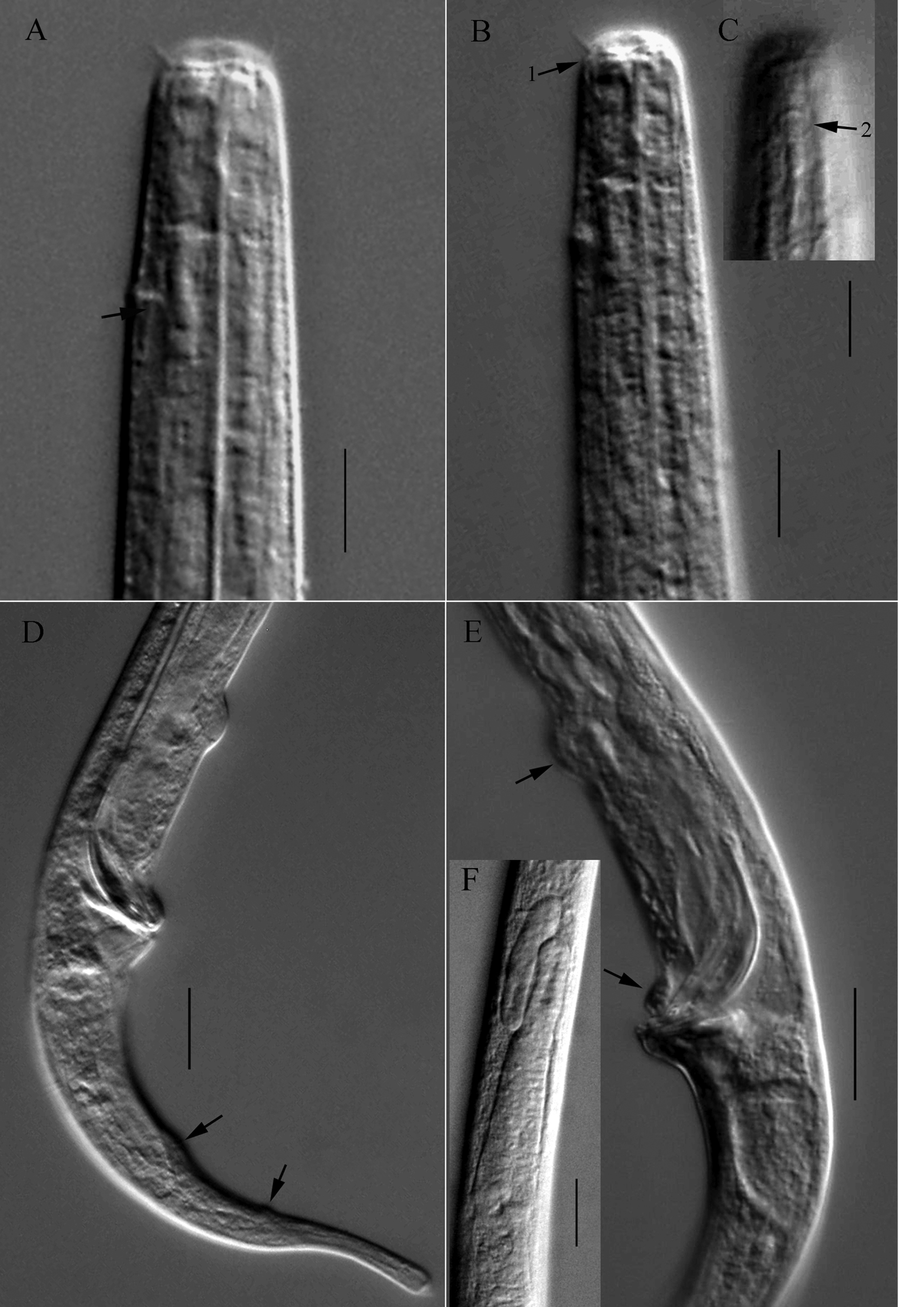
Microscopic images of Halomonhystera zhangi sp. nov. A anterior end of holotype, showing anterior setae, renette ampulla (arrow) B, C anterior end of holotype, showing buccal cavity, cephalic setae, excretory pore (arrow 1) and amphidial fovea (arrow 2) D posterior end of holotype, showing gubernaculum and caudal papillae (arrows) E cloacal region of paratype 1, showing spicule and precloacal papillae (arrows) F anterior portion of testis of holotype. Scale bars: 10 μm (A–E); 20 μm (F). |