|
||
|
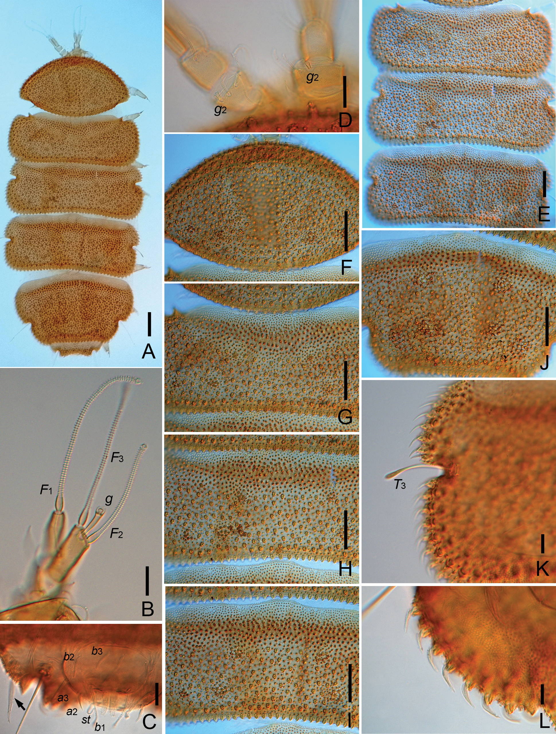
Eurypauropus japonicus Hagino & Scheller, 1985 (Chinese specimen) A habitus, tergal view, on slide B right antenna, tergal view C posterior corner of tergite VI, pygidium and anal plate (arrow indicates the big spine), sternal view D antennal segment III, showing globulus g2 E Tergite II–IV F tergites I G tergite II, middle part H tergite III, middle part I tergite IV, middle part J tergite V K tergite IV, left side, showing the marginal protuberances and T3 L tergite V, left side, showing marginal protuberances. Scale bars: 100 μm (A); 20 μm (B–L). |