|
||
|
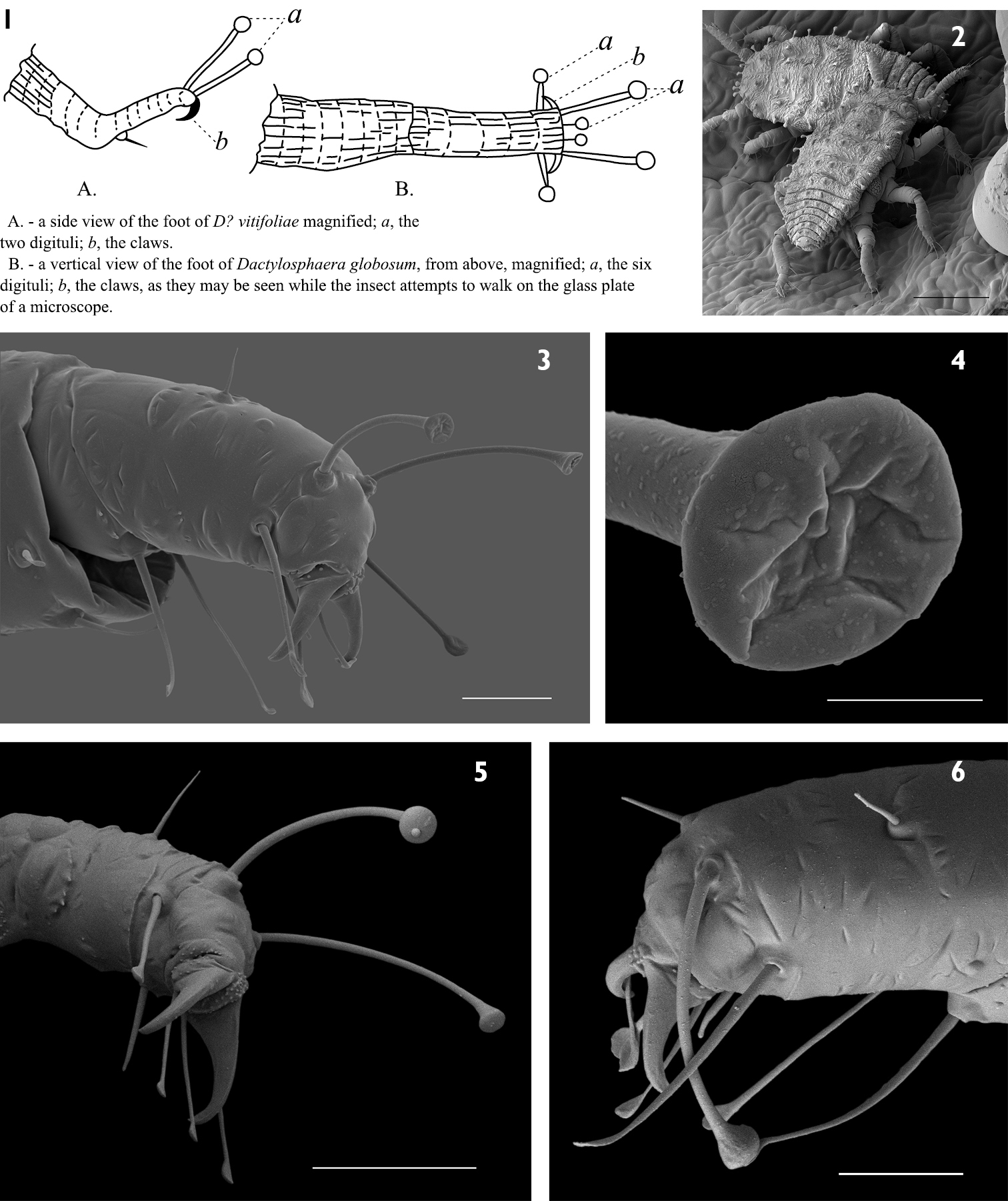
1 Reproduction of Shimer’s (1867a) original illustration indicating what he considered digitules 2 Phylloxera in situ showing leg and feet position 3 Hind leg of Phylloxera showing all digitules 4 Close up of dorsal digitule from Figure 3. Note how the ventral surface appears membranous and collapsed 5 Hind leg of Phylloxera showing dorsal digitules with expanded ventral surfaces 6 Front left leg of Phylloxera showing anterior seta is not a digitule. Scale bar 10 μm. Scale bars: (2, 6)100 μm; (3) 10 μm; (4) 2 μm; (5) 20 μm. |