- ContentsContents
- Article InfoArticle Info
- CiteCite
- MetricsMetrics
- CommentComment
- RelatedRelated
- FigsFigs
- TabsTabs
- MapMap
- TaxaTaxa
- DataData
- RefsRefs
- CitedCited
- NanopubsNanopubs
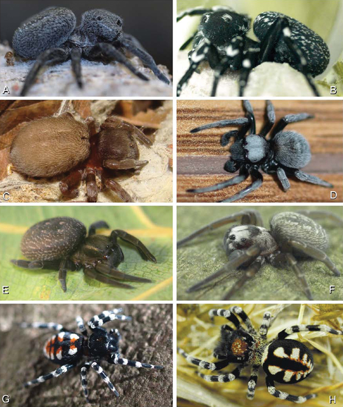
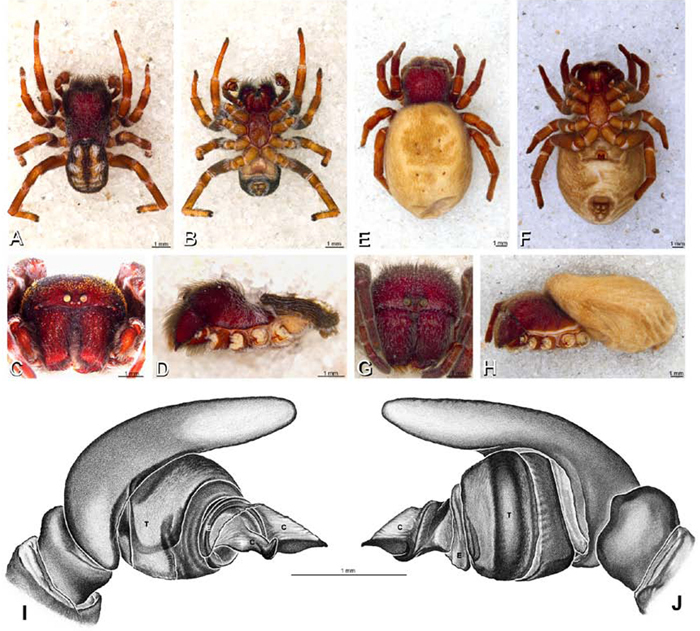
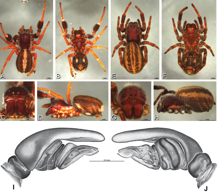
A–H Habitus of living Eresidae, photographs. A, B Adonea fimbriata A juvenile female (photo by Martin Forman) B adult male from Israel (photo by Martin Forman) C Dresserus kannemeyeri, adult female from Ndumo Game Reserve, South Africa (photo Stanislav Macík) D Dresserus sp., adult malefrom Namibia, between the towns Aus and Helmeringhausen (26°13.049'S, 16°36.063'E; photo by Martin Forman) E, F Gandanameno sp. E subadult female from Cape Town, South Africa (Stanislav Macík) F adult male from Anysberg Nature Reserve, Western Cape Province, South Africa (photo Martin Forman) G, H adult male Loureedia annulipes; G from Tel Krayot, Israel (photo by Martin Forman) H from Arad, Israel (photo by Martin Forman).
A–H Habitus of living Eresidae, photographs. A, B Eresus kollari A adult female from Hungary (photo by Tamás Szűts) B adult male from Kadaň, Czechia, (photo by Pavel Krásenský) C, E Eresus walckenaeri; C adult female from Greece (photo by Sergio Henriques) D adult male from Mihas, Greece (photo by Martin Forman) E juvenile female F Seothyra sp., juvenile female, from Brandberg, Namibia (photo by Martin Forman) G, H Paradonea variegata (photos by Martin Forman) G juvenile female from Betta, Namibia H adult male from Homeb, Namibia.
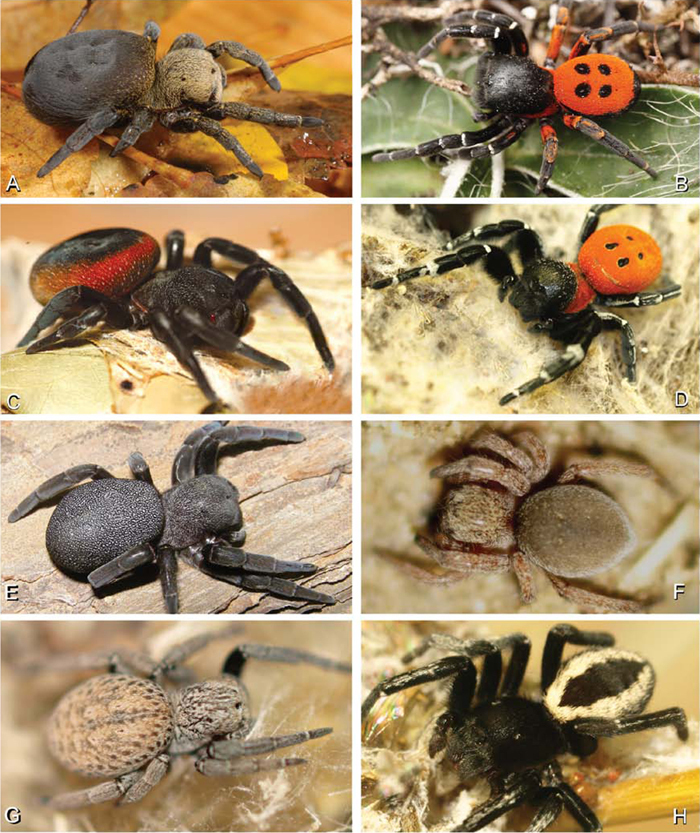
A–I Habitus of living Stegodyphus, photographs. A–C Stegodyphus lineatus A adult female from Hurghada, Egypt B adult female from Negev desert, Israel (photo by Rudolf Macek) C adult female from Shoam, Israel (photo by Amir Weinstein) D juvenile Stegodyphus tibialis feeding on their mother, Dali, China (photo by Yang Zi-Zhong) E Stegodyphus mimosarum, male (black arrow), females and a kleptoparasite Archeodictyna (white arrow) F Stegodyphus mimosarum, mass attack on a carabid G a female Stegodyphus dumicola feeding her offsprings H a pompilid wasp larva feeds on a female Stegodyphus dumicola (photos E–H by Teresa Meikle) I Stegodyphus sp. female from ShweSettaw Wildlife Reservation, Myanmar (photo by Dong Lin).
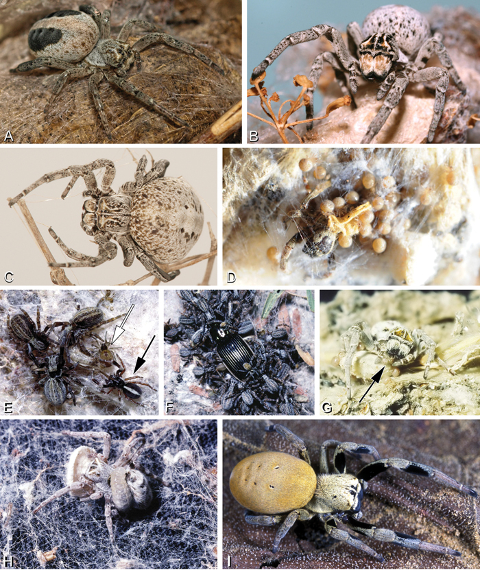
A–L Retreats, webs, and habitus of living Eresidae, photographs. A Adonea fimbriata retreat on the ground from Sede Boqer, Israel (photo by Efrat Gavish-Regev) B Dresserus sp. retreat in the grass (photo by Charles Haddad) C Eresus walckenaeri retreat of juvenile from Ioannina, Greece (photo by Siegfried Huber) D–E Gandanameno sp. from west of Helmeringhausen Namibia (photos by Martin Forman) D Retreat on Acacia E female, with egg sac and various prey remnants F Gandanameno sp. femalefrom Amanzi Game Reserve, South Africa (photo by Tamás Szűts) G, H Seothyra sp. from Namibia G retreat under sand, showing the antelope track pattern H specimen and the exposed retreat (photos E–H, J, L by Teresa Meikle) I Loureedia annulipes, burrow from Sede Boqer, Israel (photo by Efrat Gavish-Regev) J–L Stegodyphus retreats J Stegodyphus dumicola from Spioenkop, South Africa K Stegodyphus lineatus from Tel-Hadid, Israel (photo by Amir Weinstein) L Stegodyphus mimosarum from Spioenkop, South Africa.
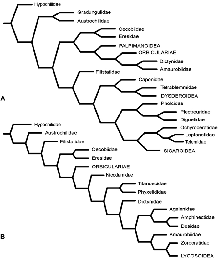
A–B Historical hypotheses of the phylogenetic position of Eresidae. A simplified cladogram from Platnick et al. (1991: 68, fig. 311) B simplified cladogram from Griswold et al. (1999: 58, fig. 1). Terminals merged into family level (normal type) or higher level (all capital type) taxa.
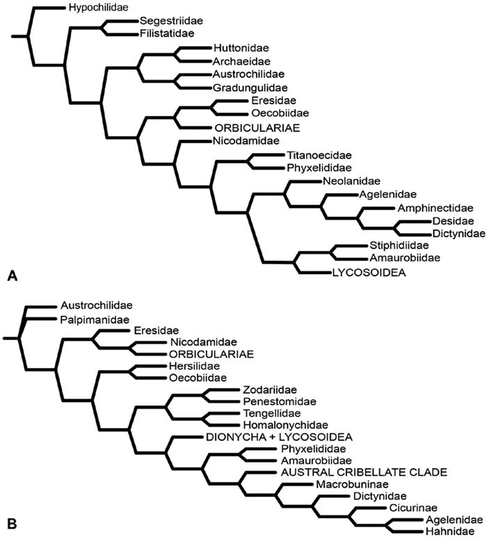
A–B Historical hypotheses of the phylogenetic position of Eresidae. A simplified implied weights cladogram (K=6, fit=115.93, L=488) from Griswold et al. (2005: 316, fig. 219) B simplified Bayesian tree from Miller et al. (2010a: 792, fig. 3). Terminals merged into family level (normal type) or higher level (all capital type) taxa.
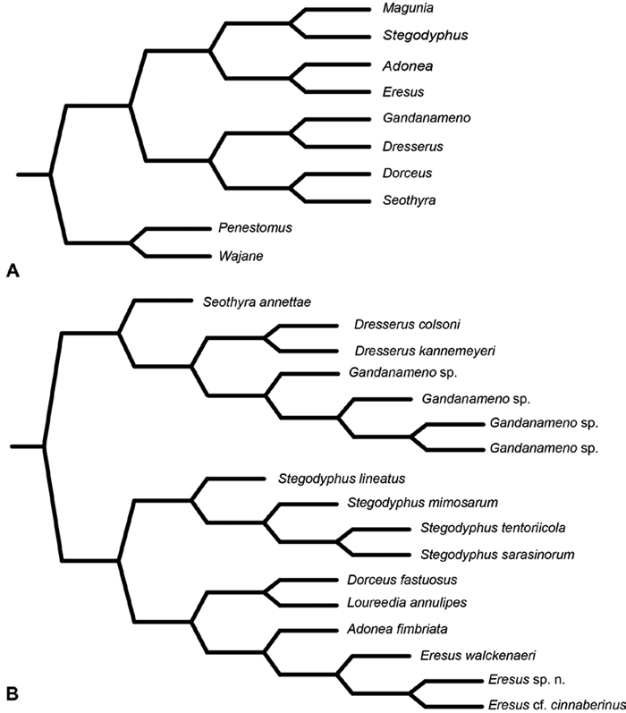
A–B Historical phylogenetic hypotheses of relationships among Eresidae. A tree from Lehtinen (1967: 387, fig. 13). Magunia was synonymized with Stegodyphus by Kraus and Kraus (1988); Wajane was synonymized with Penestomus and removed from Eresidae by Miller et al. (2010a; 2010b) B relationships among Eresidae based on molecular phylogenetic analysis of Miller et al. (2010a: 792, modified fig. 3). “Stegodyphus" annulipes relabeled Loureedia annulipes to reflect a nomenclatural change proposed in this work and Gandanameno species epithets removed to reflect increasing uncertainty about species limits and identity in this genus.
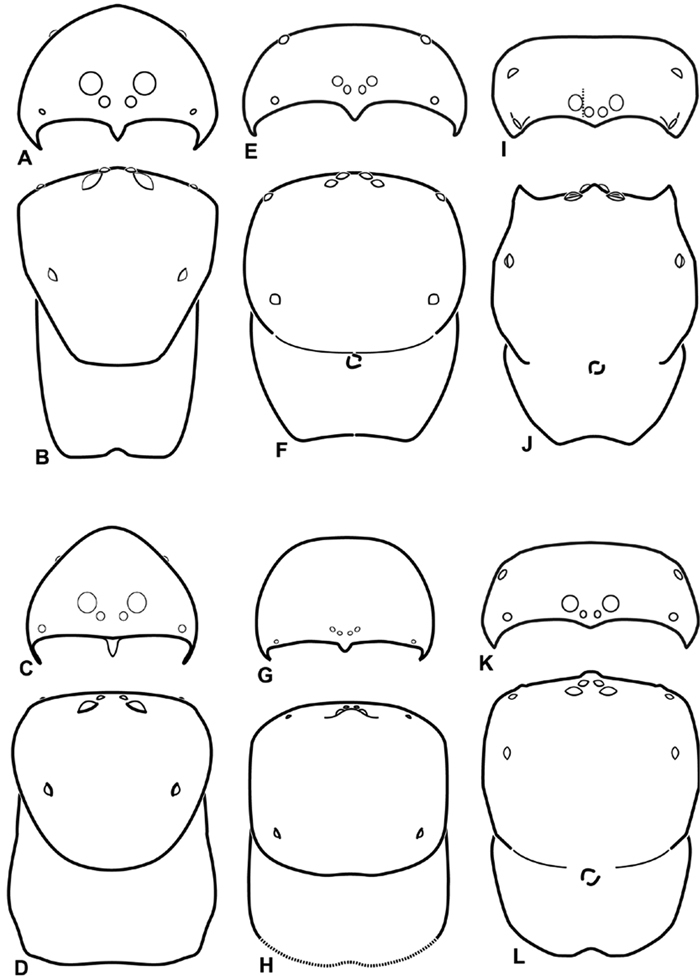
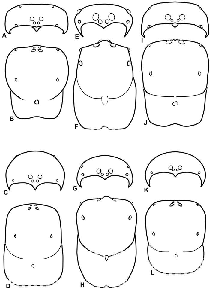
A–L Schematic illustrations of the carapace of assorted eresids A–D Adonea fimbriata E–H Dorceus fastuosus I–L Dresserus sp. A–B, E–F, I–J male C–D, G–H, K–L female A, C, E, G, I, K anterior view B, D, F, H, J, L dorsal view. Dashed line in I drawn tangential to the mesal margin of the PME does not intersect with the AME indicating median eyes separated on vertical axis. Dashed lines at posterior of carapace indicate uncertainty. Not to scale.
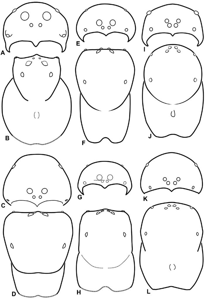
A–L Schematic illustrations of the carapace of assorted eresids. A–D Eresus kollari E–H Gandanameno sp. I–L Loureedia annulipes A–B, E–F, I–J male C–D, G–H, K–L female A, C, E, G, I, K anterior view B, D, F, H, J, L dorsal view. Dashed lines at posterior of carapace indicate uncertainty. Not to scale.
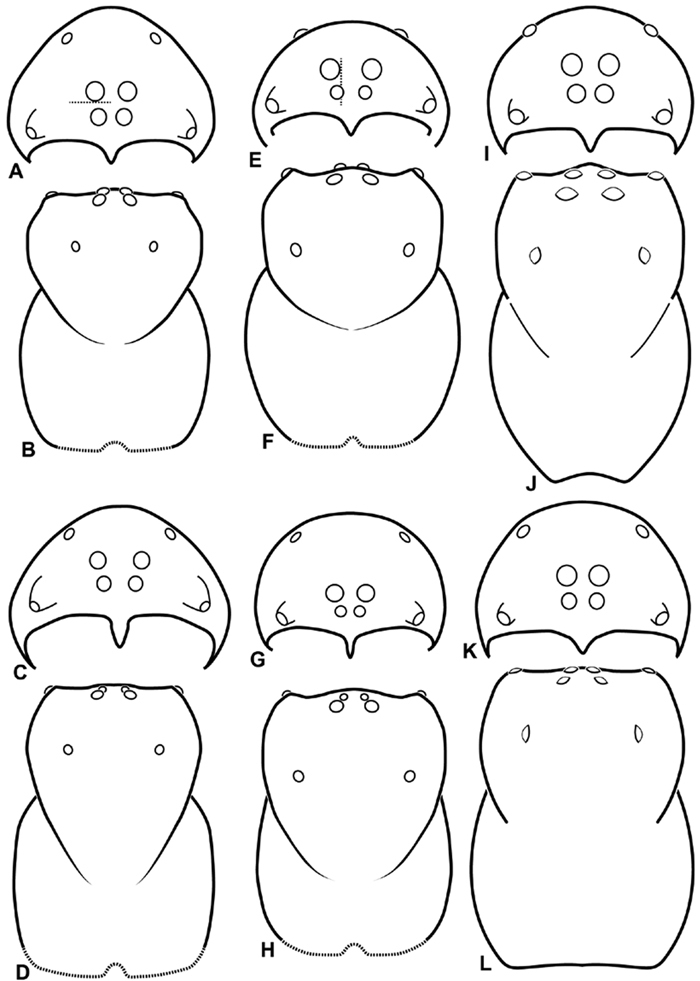
A–L Schematic illustrations of the carapace of assorted eresids. A–B Paradonea striatipes C–D Paradonea splendens E–H Paradonea variegata I–L Seothyra henscheli A–D, E–F, I–J male G–H, K–L female. A, C, E, G, I, K anterior view B, D, F, H, J, L dorsal view G illustrates example of median eyes overlapping on horizontal axis. Dashed lines at posterior of carapace indicate uncertainty. Not to scale.
A–L Schematic illustrations of the carapace of assorted Stegodyphus species. A–D Stegodyphus lineatus E–H Stegodyphus mimosarum I–L Stegodyphus sarasinorum. A–B, E–F, I–J male C–D, G–H, K–L female A, C, E, G, I, K anterior view B, D, F, H, J, L dorsal view A illustrates example of median eyes separated on horizontal axis; E illustrates example of median eyes overlapping on vertical axis. Dashed lines at posterior of carapace indicate uncertainty. Not to scale.
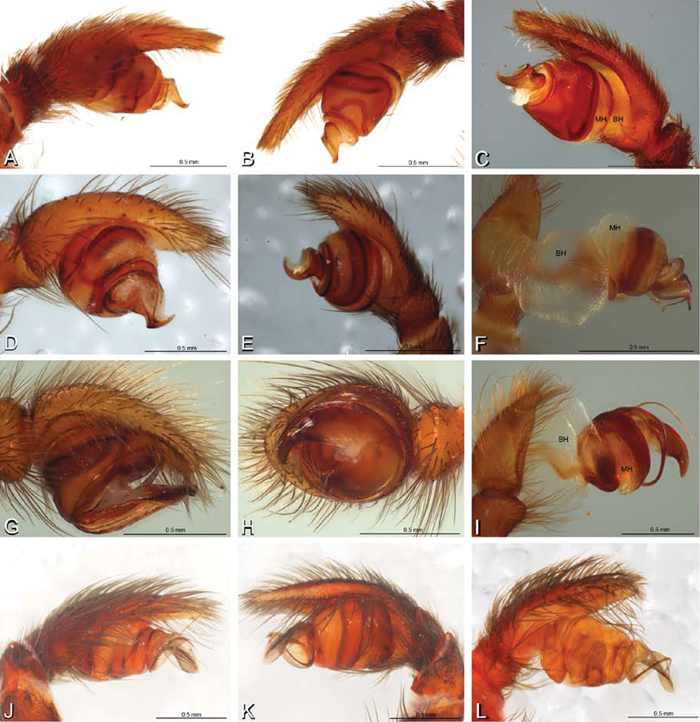
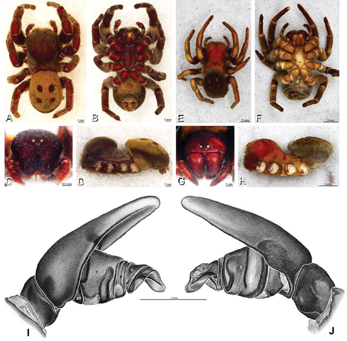
A–L Left male palpi of eresid species, photomicrographs. A–C Adonea fimbriata from Algeria-Morocco (MR012, MR) D–F Dorceus fastuosus from Mashabin Sand Dunes, Israel (MR006, HUJ) G–I Dresserus sp. from Manga Forest Reserve, Tanzania J–L Eresus walckenaeri from Leptokaryas, Greece (MR020, MR) A, D, G, J prolateral view B, E, K retrolateral view H ventral view C, F, I, L expanded palp. BH basal haematodocha MH median haematodocha.
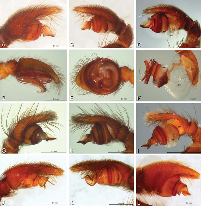
A–L Left male palpi of eresid species, photomicrographs. A–C Eresus kollari from res. Radotinske udoli, Czechia (MR007, MR) D–F Gandanameno sp. from Van Riebeeck Park, Western Cape, South Africa (CASENT 9023763, CAS) G–I Loureedia annulipes from Haluqim Ridge, Israel (PET03, MR) J, K Paradonea striatipes from Otjivasandu (NMN), Namibia L Paradonea splendens from Sunnyside, South Africa (C1076, SAM) A, D, G, J, L prolateral view B, H, K retrolateral view E ventral view C, F, I expanded palp. BH basal haematodocha MH median haematodocha.
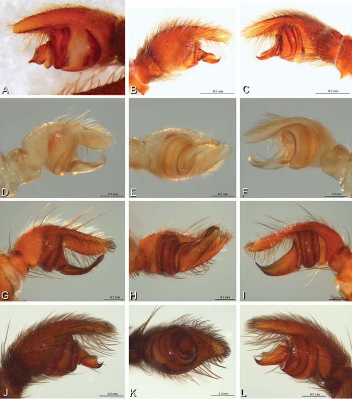
A–L Left male palpi of Paradonea species, photomicrographs. A Paradonea splendens from Sunnyside, South Africa (C1076, SAM) B, C Paradonea variegata from Breekkierie Dunes, Northern Cape, South Africa (C1062, SAM) D–I Paradonea parva D–F holotype from junction of Marico and Crocodile Rivers, South Africa (B3701, SAM) G–I from 4 km N of Hopetown, Northern Cape, South Africa (AcAT 97/988, NCA) J–L Paradonea presleyi sp. n. holotype from Falcon College, Zimbabwe (CASENT 9039236, CAS) A, C, F, I, L retrolateral view B, D, G, J prolateral view E, H, K ventral view.
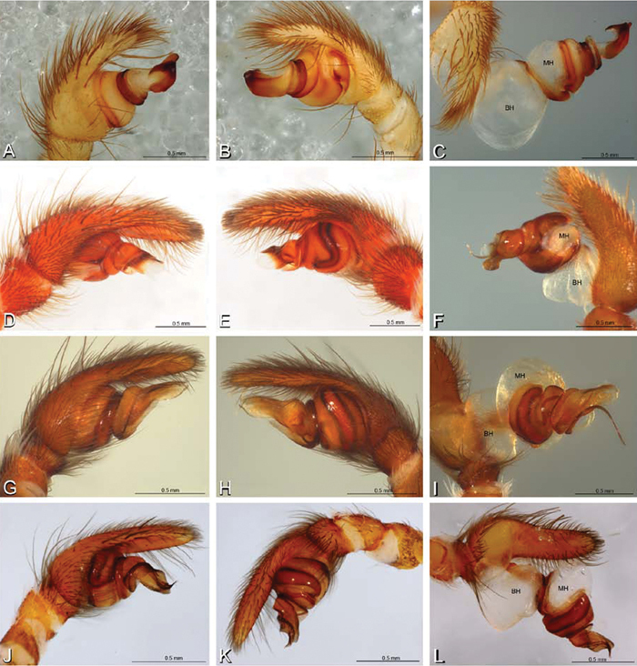
A–L Left male palpi of eresid species, photomicrographs. A–C Seothyra henscheli from Gobabeb Station, Namibia (SMN 40828, NMN) D, F Stegodyphus lineatus D–E from Negev, Israel (MR) F from Nengrahar, Afghanistan (MR010, MR) G–I Stegodyphus mimosarum from Forêt d'Analalava, Fianarantsoa, Madagascar (CASENT 9015950, CAS) J–L Stegodyphus sarasinorum from 7.5 km E PwintPhyu, Magway Division, Myanmar (CASENT 9019370, CAS) A, D, G, J prolateral view B, E, H, K retrolateral view C, F, I, L expanded palp. BH basal haematodocha MH median haematodocha.

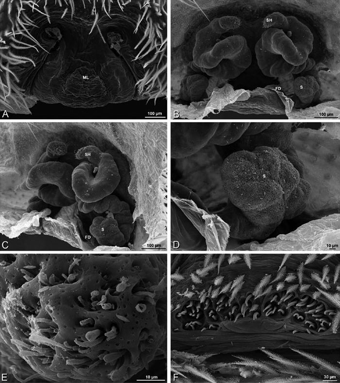
A–L Epigyna of eresid species, photomicrographs. A, D Adonea fimbriata; A from Mehav Am village, Israel (MR003, MR) D from Wadi Mashash, Israel (MR013, HUJ) B, E Dorceus fastuosus from Mashabim sand dunes, Israel (MR002, MR) C, F Dresserus sp. from Klein Kariba, South Africa (CASENT 9025745, CAS) G, J Eresus walckenaeri from 5 km south of Monemvasia, Lakonia, Greece (ZMUC 00012903, ZMUC) H, K Eresus kollari from res. Radotinske udoli, Czechia (MR016, MR) I, L Eresus sandaliatus from SE of Silkeborg, Denmark (CASENT 9039243, CAS) A–C, G–I ventral viewD–F, J–L dorsal view, cleared. CD copulatory duct ML median lobe S spermatheca SH spermathecal head.
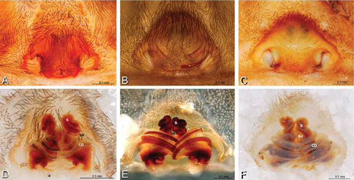
A–F Epigyna of Gandanameno sp., photomicrographs. A, D from Iringa, Tanzania (ZMUC 19970517, ZMUC) B, E from Kommetjie, Western Cape, South Africa (CASENT 9039241, CAS), note broken embolus left in female reproductive system C, F from Port Elizabeth, South Africa (port-3325, ZMHB) A–C ventral view D–F dorsal view, cleared. CD copulatory duct S spermatheca SH spermathecal head.
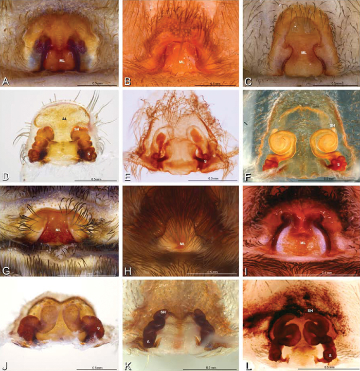
A–L Epigyna of eresid species, photomicrographs. A, D Loureedia annulipes from Wadi Mashash, Negev, Israel (MR019, MR) B, E Paradonea variegata from Steinkopf, Northern Cape, South Africa (ZMB 26964, ZMHB) C, F Seothyra henscheli; C from Kuiseb River, Gobabeb, Namibia (SMN 46627, NMN) F from Sout Rivier, Namibia (CASENT 9039242, CAS) G, J Stegodyphus lineatus from Belkis, near Birecor, Turkey (MR015, MR) H, K Stegodyphus mimosarum H from Forêt d'Analalava, Fianarantsoa, Madagascar (CASENT 9015950, CAS) K from Réserve Spéciale de Cap Sainte Marie, Toliara, Madagascar (CASENT 9012844, CAS) I, L Stegodyphus sarasinorum from 7.5 km E PwintPhyu, Magway Division, Myanmar (CASENT 9019370, CAS) A–C, G–I ventral view D–F, J–L dorsal view, cleared. AL anterior lobe ML median lobe S spermatheca SH spermathecal head.
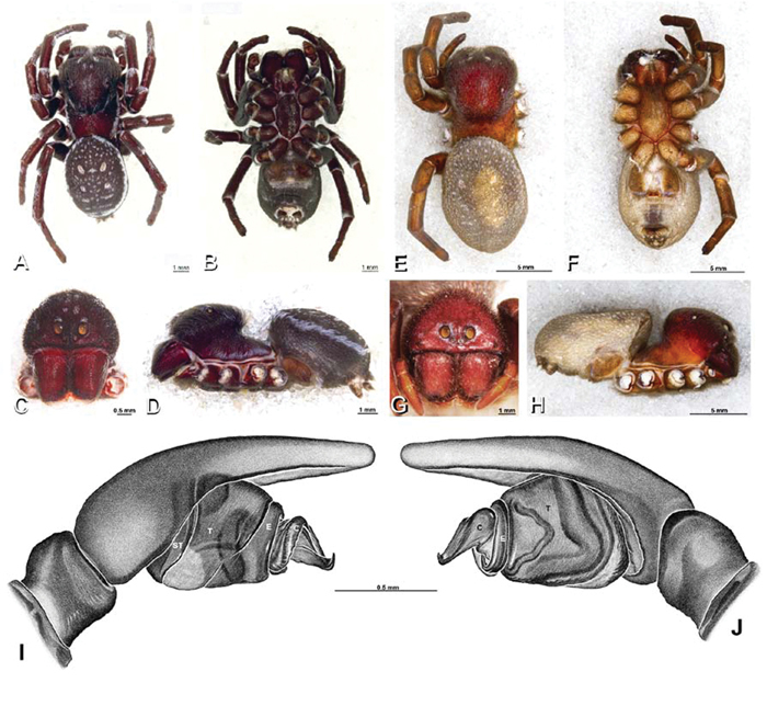
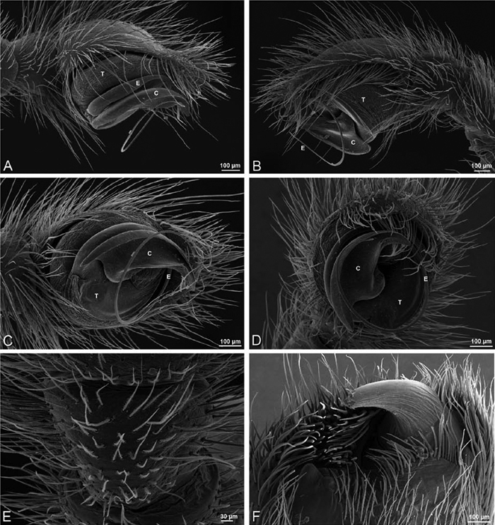
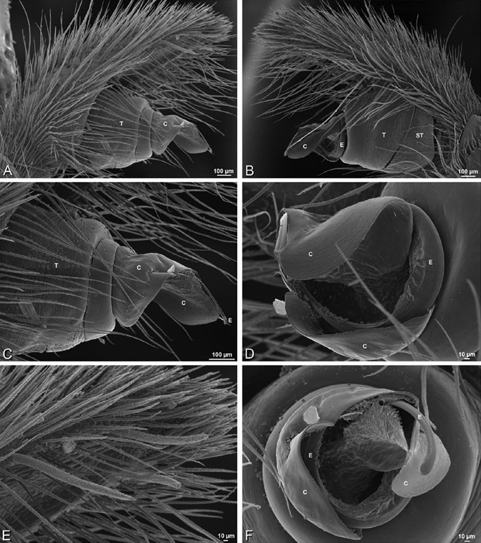
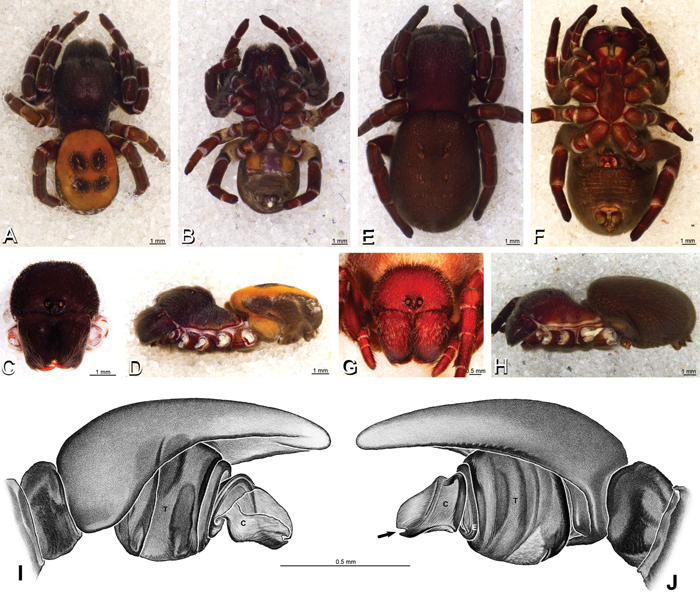
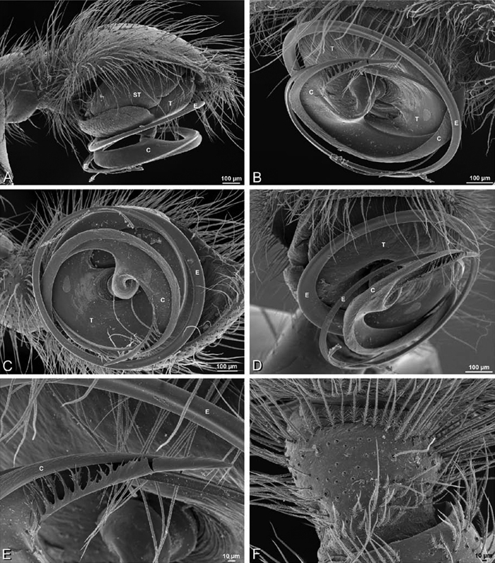
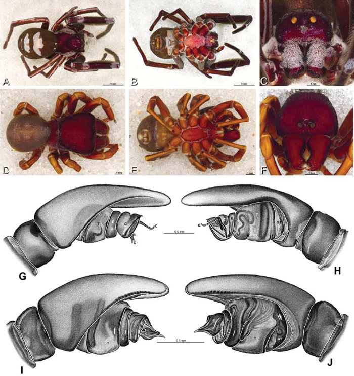
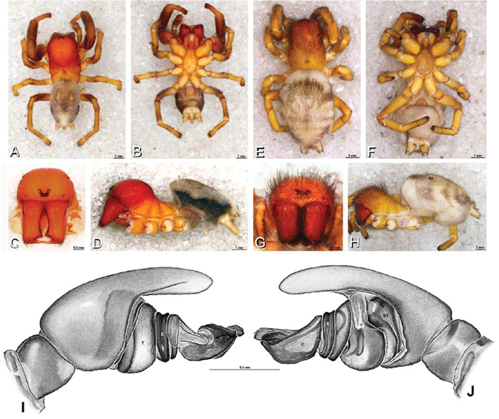
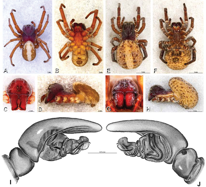
A–J Adonea fimbriata. A–D, I–J male from Algeria-Morocco (MR012, MR) E–H female from Mehav Am village, Israel (MR003, MR) A–D habitus of male, photomicrographs E–H habitus of female photomicrographs I, J illustrations of left male palp A, E dorsal view B, F ventral view C, G anterior view. D, H lateral view I prolateral view J retrolateral view. C conductor E embolus ST subtegulum T tegulum.

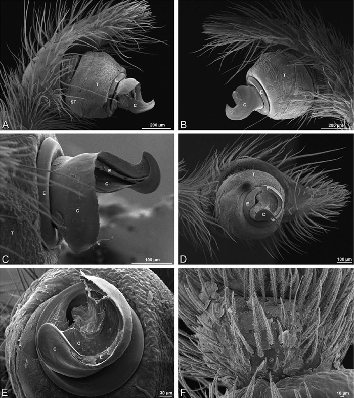
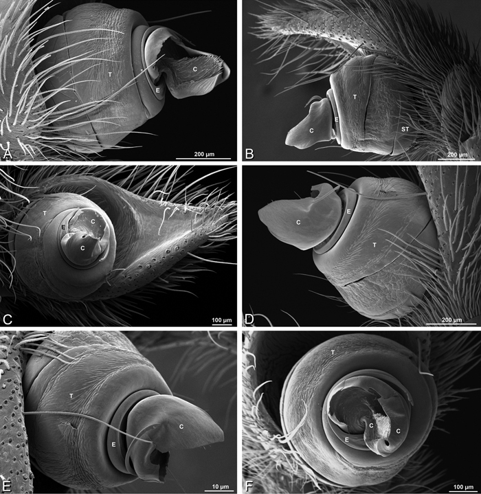
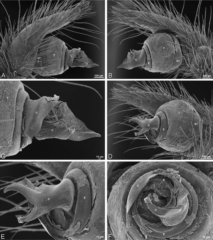
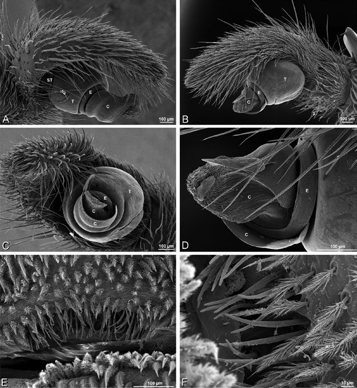
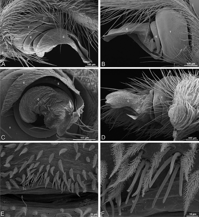
A–F Adonea fimbriata from Algeria-Morocco (MR012, MR), scanning electron micrographs of right male palp, images reversed to appear as left palp. A prolateral view B retrolateral view C detail of embolic division, prolateral view D detail of embolic division, retrolateral view E ventral view F apical view. C conductor E embolus ST subtegulum T tegulum.
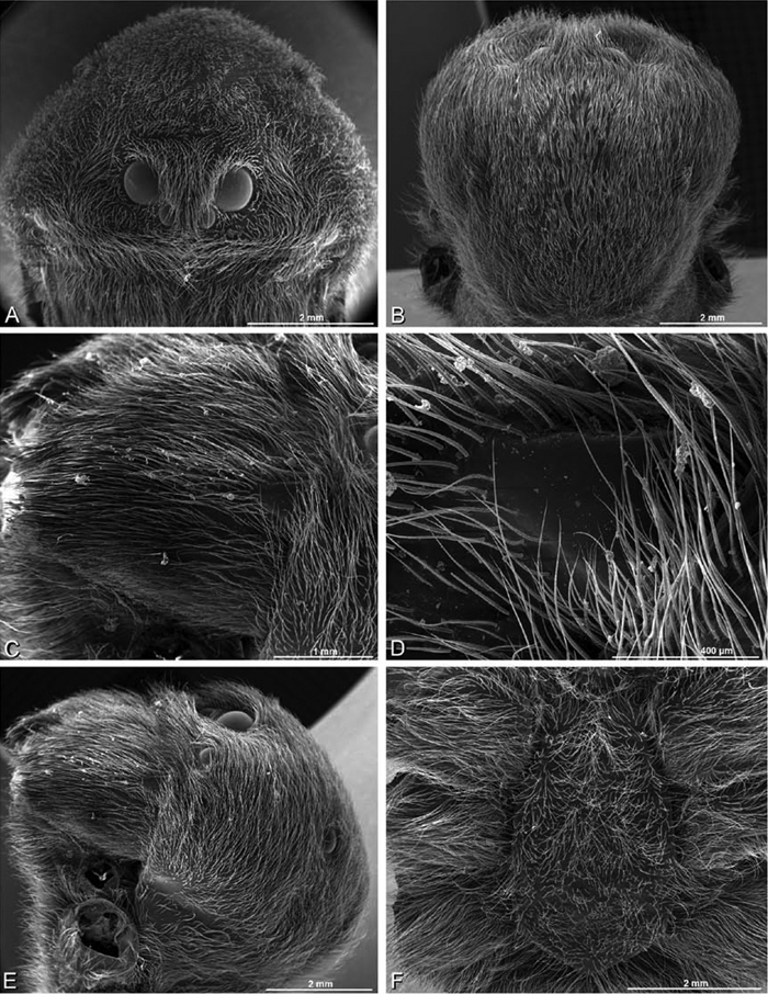
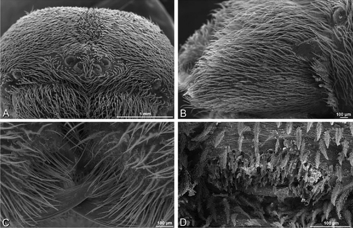
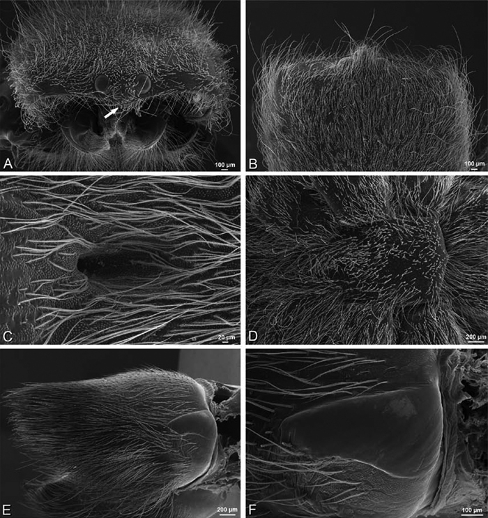
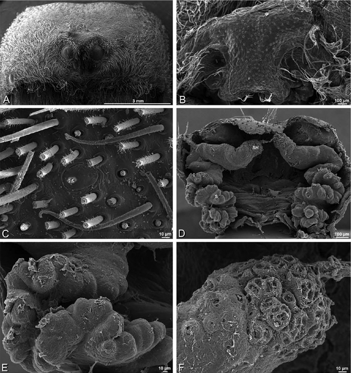
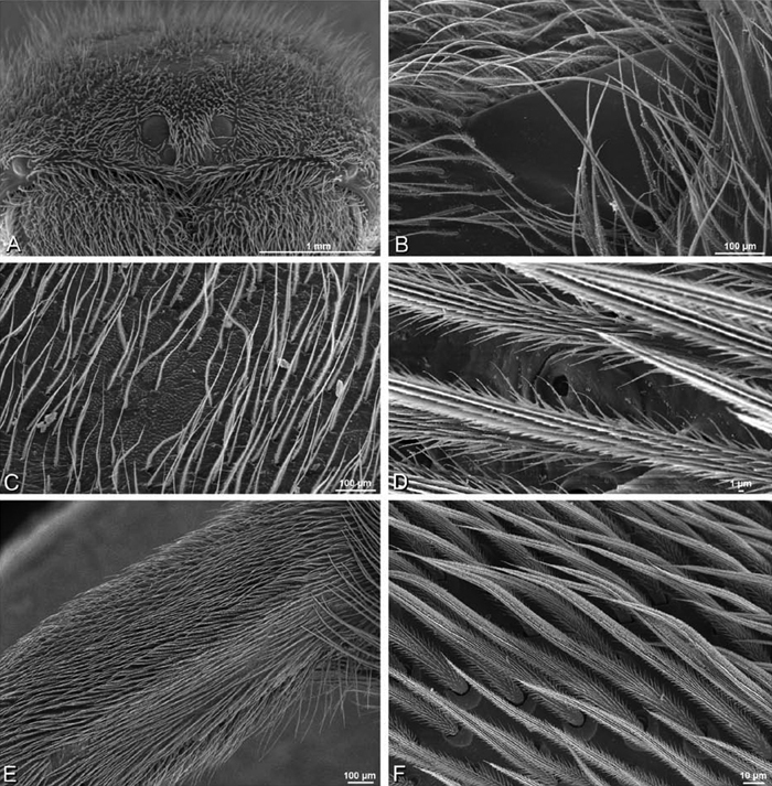
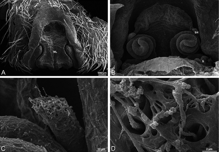
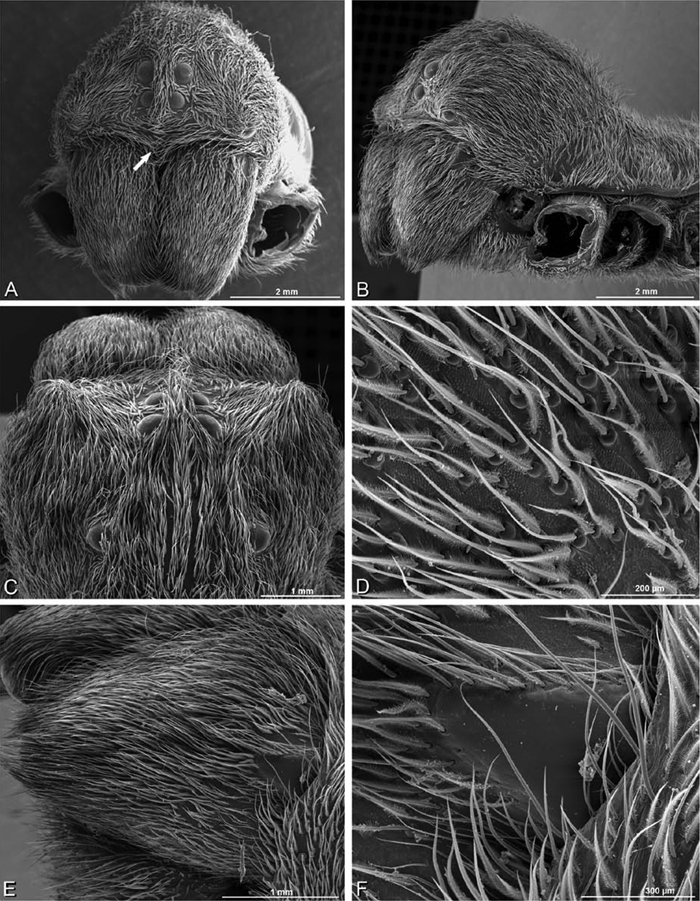
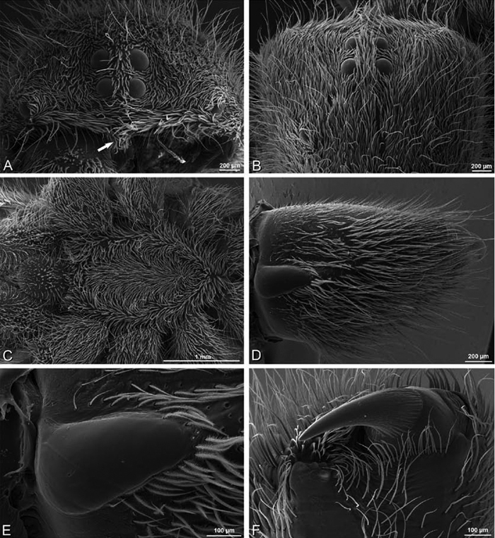
A–F Adonea fimbriata from Mehav Am village, Israel (MR003, MR), scanning electron micrographs of female prosoma. A anterior view B dorsal view C left chelicerae, lateral view D left cheliceral boss E lateral view F sternum and coxae, ventral view
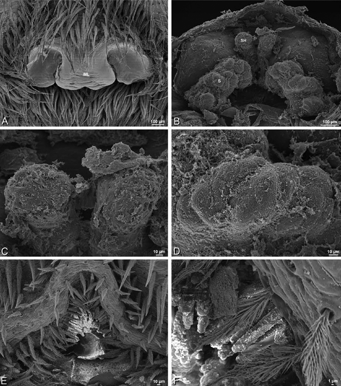
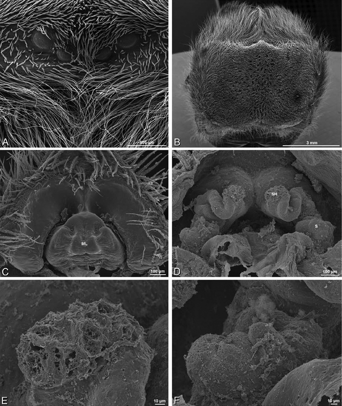
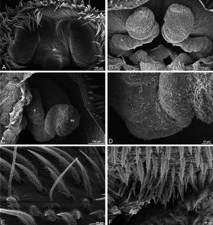
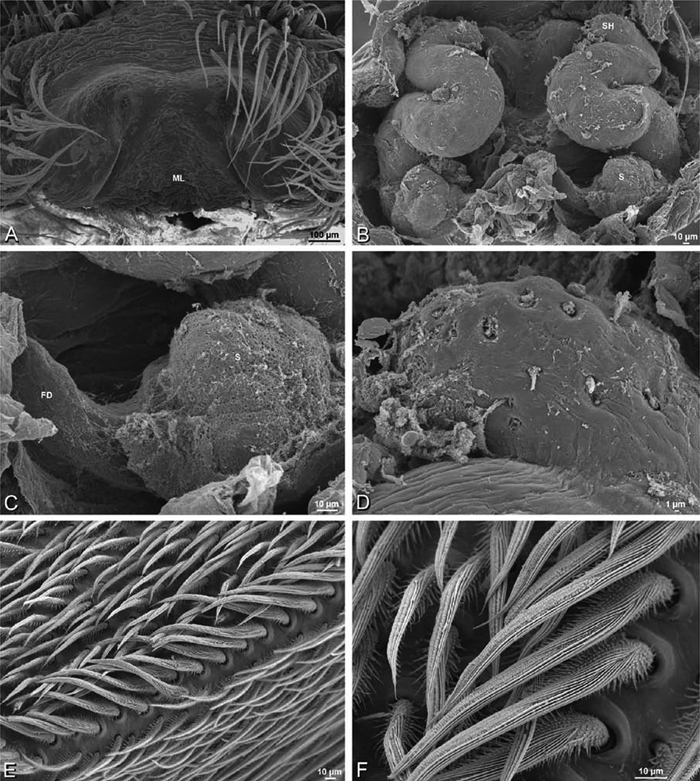
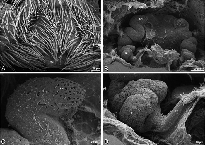
A–F Adonea fimbriata, scanning electron micrographs. A female from Mehav Am village, Israel (MR003, MR) B–D female from Wadi Mashash, Israel (MR013, HUJ) E, F male from Algeria-Morocco (MR012, MR) A–D vulva E, F epiandrous region A epigynum, ventral view B cleared vulva, dorsal view C detail, spermathecal heads D detail, right spermatheca E epiandrous region F detail of epiandrous gland spigots. ML median lobe S spermatheca SH spermathecal head.
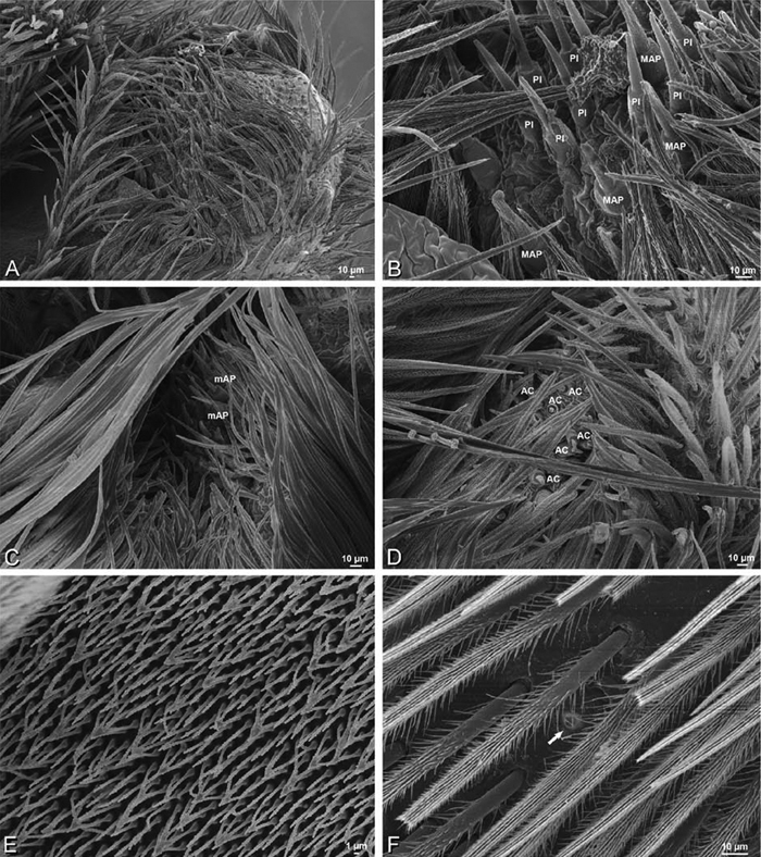
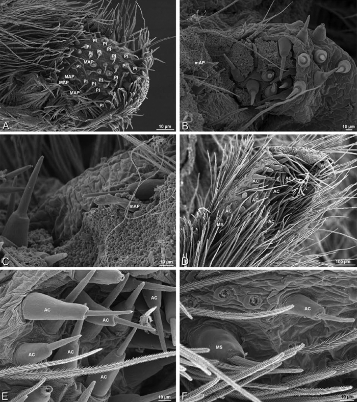
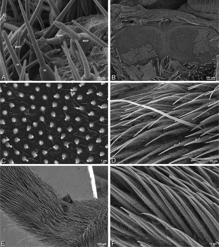
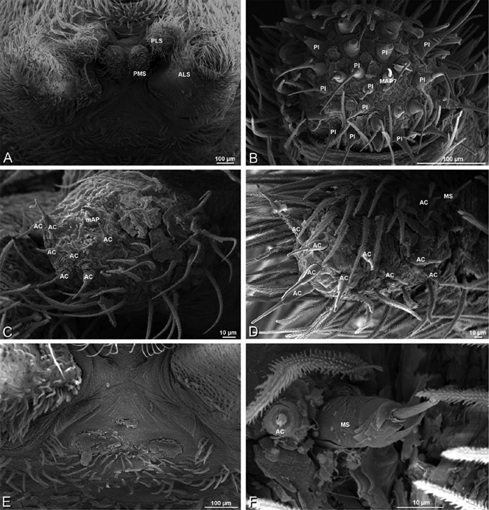
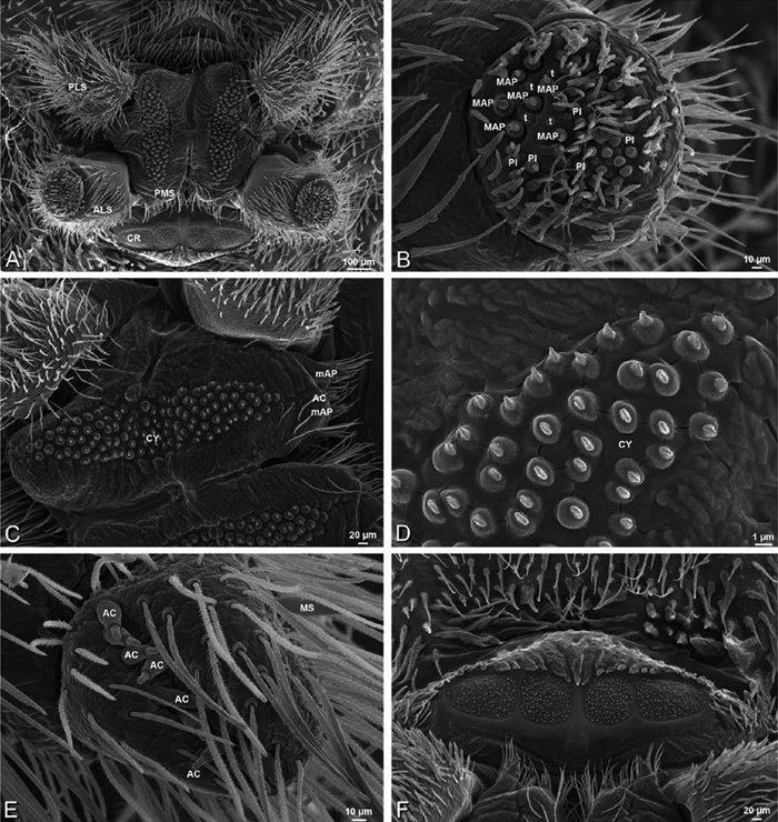
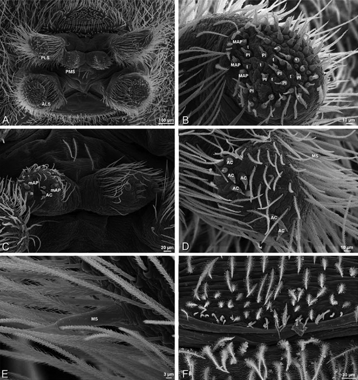
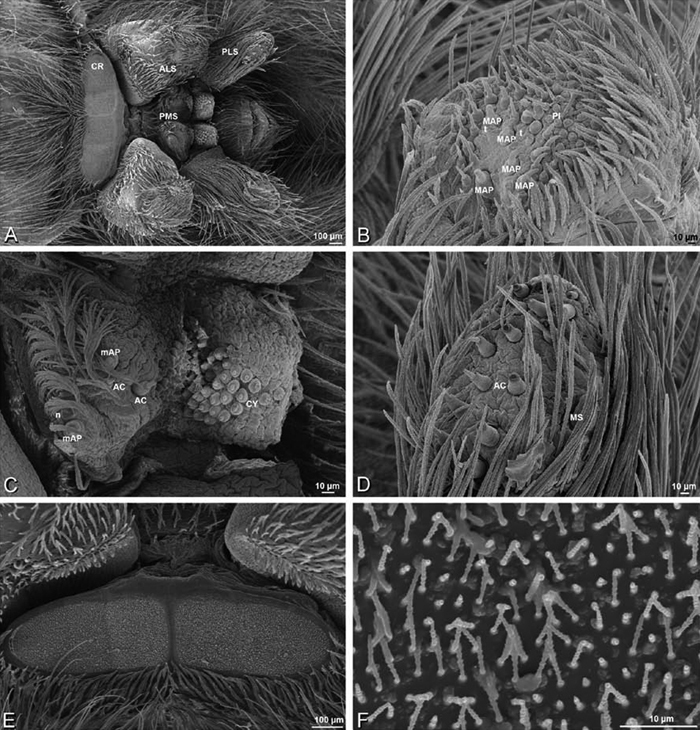
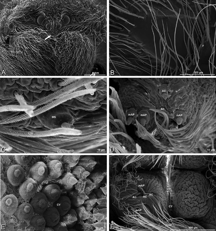
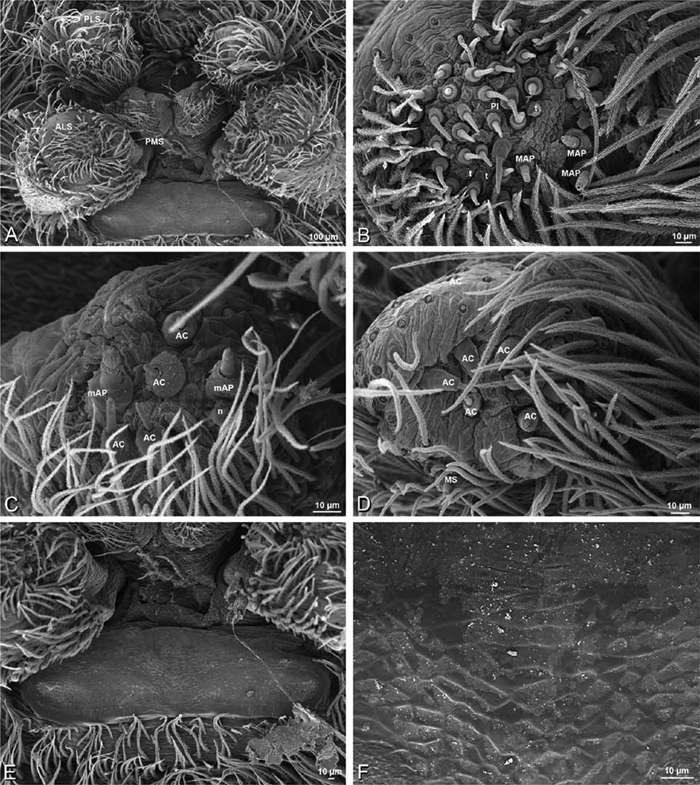
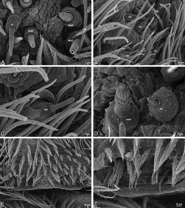
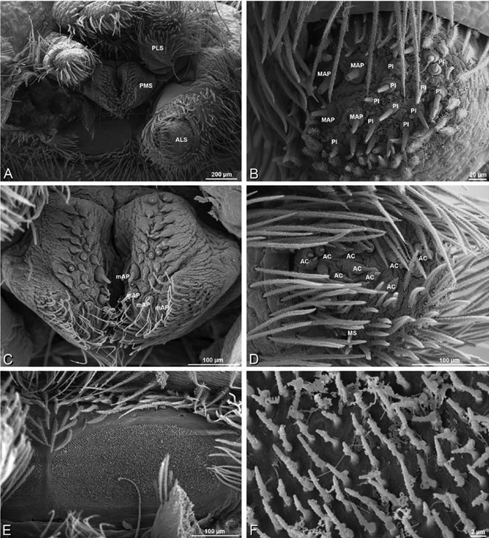
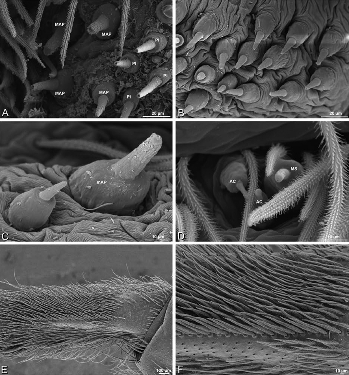
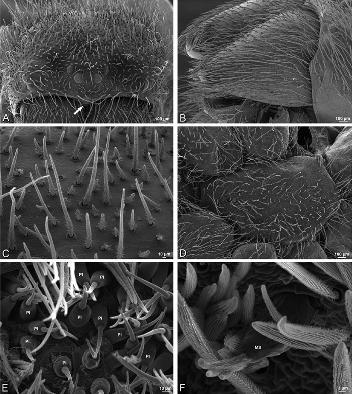
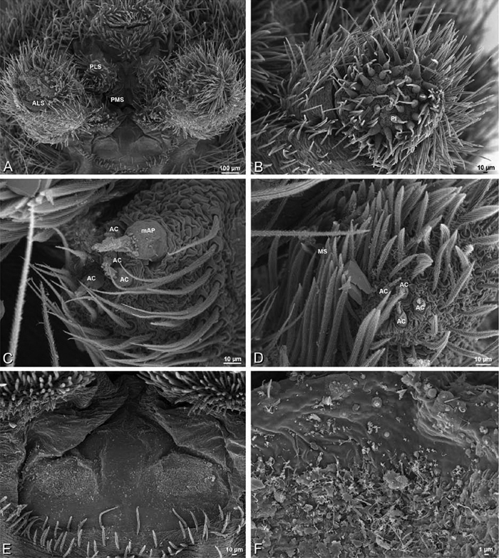
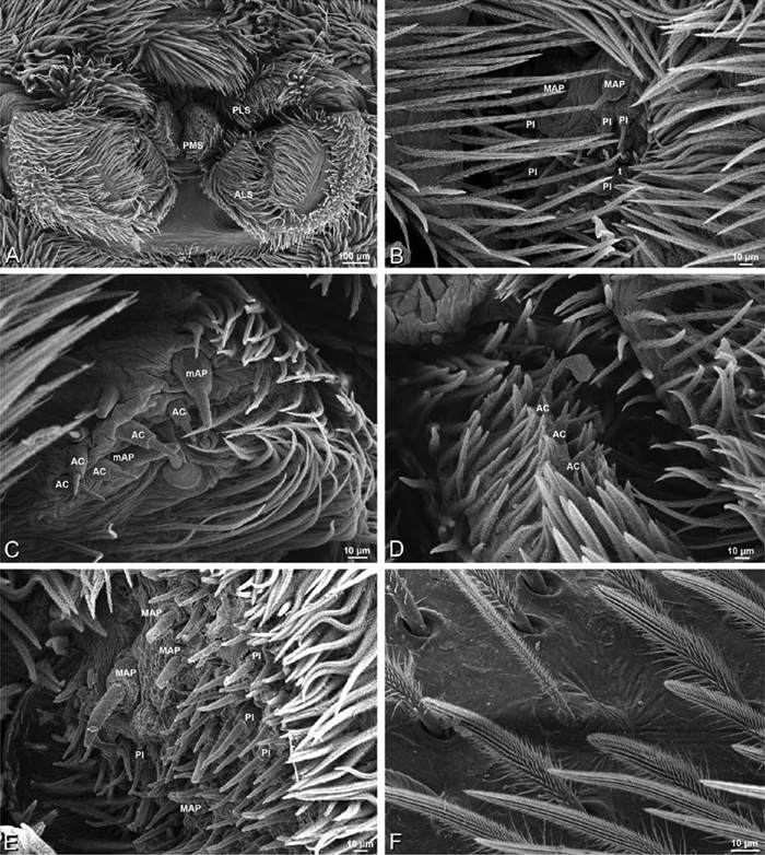
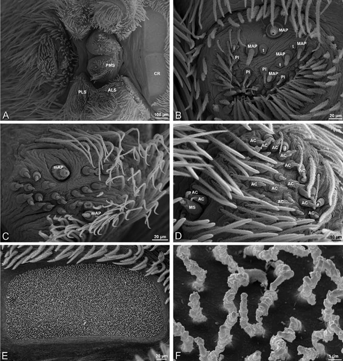
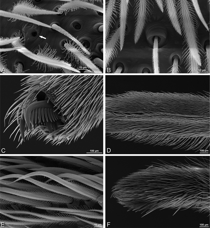
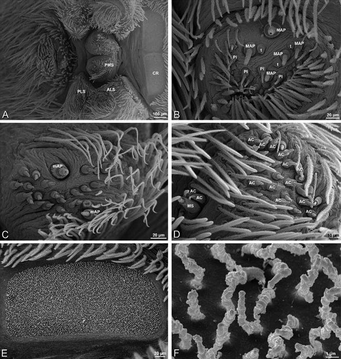
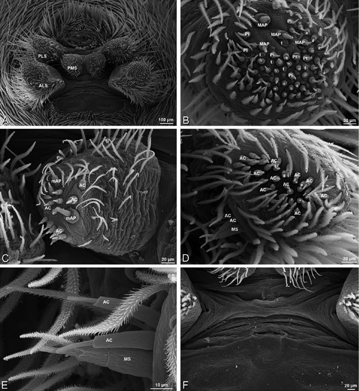
A–F Adonea fimbriata, female from Mehav Am village, Israel (MR003, MR), scanning electron micrographs of spinnerets. A right ALS B detail of spigots on right ALS C PMS D right PLS E cribellar spigots F arrow indicating tarsal organ, left leg I. Unlabeled spigots in C thought to be a mixture of aciniform gland spigots and cylindrical gland spigots. AC aciniform gland spigot MAP major ampullate gland spigot mAP minor ampullate gland spigot PI piriform gland spigot.
A–F Adonea fimbriata, male from Algeria-Morocco (MR012, MR), scanning electron micrographs of spinnerets. A overview B right ALS C right PMS D right PLS E aciniform field on right PLS F modified spigot on right PLS. AC aciniform gland spigot ALS anterior lateral spinneret MAP major ampullate gland spigot mAP minor ampullate gland spigot MS modified spigot PI piriform gland spigot PLS posterior lateral spinneret PMS posterior median spinneret.
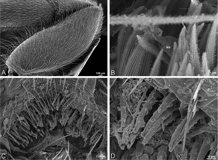
A–F Adonea fimbriata, scanning electron micrographs. A–C male from Algeria-Morocco (MR012, MR) D–F female from Mehav Am village, Israel (MR003, MR) A–C spinnerets and vestigial cribellum. D–F legs of female A detail of spigots on right male ALS B vestigial cribellum C detail of vestigial cribellum D trichobothrium, left metatarsus I E calamistrum, right metatarsus IV F detail, calamistrum seta, right metatarsus IV. MAP major ampullate gland spigot.
A–J Dorceus fastuosus. A–D, I–J male from Mashabin Sand Dunes, Israel (MR006, HUJ) E–H female from Mashabim sand dunes, Israel (MR002, MR) A–D habitus of male, photomicrographs E–H habitus of female, photomicrographs J, K illustrations of left male palp. A, E dorsal view B, F ventral view C, G anterior view D, H lateral view. I prolateral view. J retrolateral view. C conductor E embolus ST subtegulum T tegulum.
A–F Dorceus fastuosus from Mashabin Sand Dunes, Israel (MR006, HUJ), scanning electron micrographs of left male palp. A prolateral view B retrolateral view C detail of embolic division, prolateral view D ventral view E detail of embolic division, ventral view F palpal tibia, dorsal view. C conductor E embolus ST subtegulum T tegulum.
A–D Dorceus fastuosus, male from Mashabin sand dunes, Israel (MR006, HUJ), scanning electron micrographs. A prosoma, anterior view B left chelicerae, lateral view C chelicerae, anterior distal view showing fangs and teeth D epiandrous region.
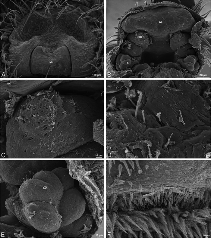
A–F Dorceus fastuosus, female from Mashabim sand dunes, Israel (MR002, MR), scanning electron micrographs. A median eye group B prosoma, dorsal C epigynum, ventral view D cleared vulva, dorsal view E detail, left spermathecal head F detail, right spermatheca. ML median lobe S spermatheca SH spermathecal head.
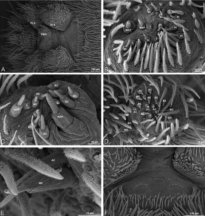
A–F Dorceus fastuosus, female from Mashabim sand dunes, Israel (MR002, MR), scanning electron micrographs of spinnerets. A left ALS B left PMS C detail of left PMS D left PLS E aciniform field on left PLS F modified spigot and flanking aciniform gland spigot on left PLS. Unlabeled spigots in B and C thought to be a mixture of aciniform gland spigots and cylindrical gland spigots. AC aciniform gland spigot MAP major ampullate gland spigot mAP minor ampullate gland spigot MS modified spigot PI piriform gland spigot.
A–F Dorceus fastuosus, female from Mashabim sand dunes, Israel (MR002, MR), scanning electron micrographs. A detail of spigots on left ALS B cribellum C detail cribellar spigots D trichobothrium, left tibia IV E calamistrum, left metatarsus IV F detail, calamistrum seta, left metatarsus IV. MAP major ampullate gland spigot PI piriform gland spigot.
A–F Dorceus fastuosus, male from Mashabin Sand Dunes, Israel (MR006, HUJ), scanning electron micrographs of spinnerets. A overview B left ALS C left PMS D left PLS E vestigial cribellum F modified spigot and flanking aciniform spigot on left PLS. AC aciniform gland spigot ALS anterior lateral spinneret MAP major ampullate gland spigot mAP minor ampullate gland spigot MS modified spigot PI piriform gland spigot PLS posterior lateral spinneret PMS posterior median spinneret.
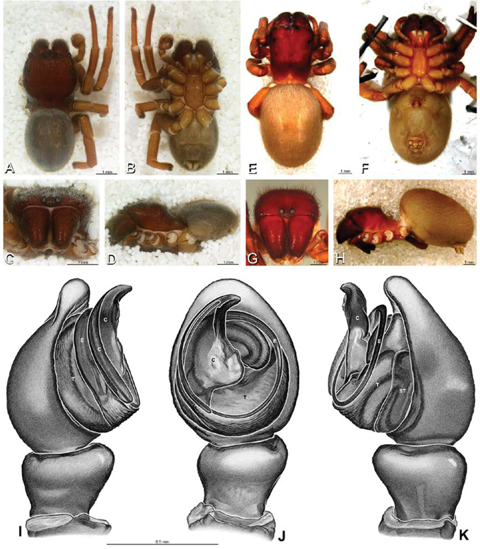
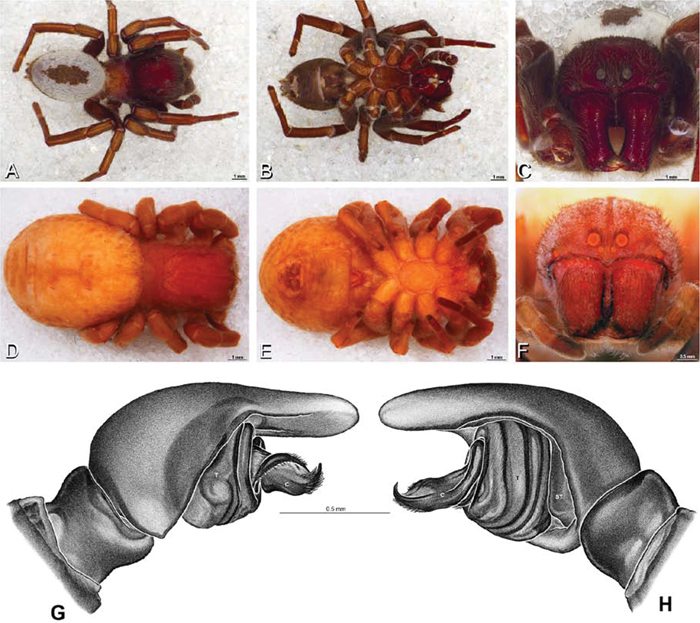
A–K Dresserus sp. A–D male from Manga Forest Reserve, Tanzania (ZMUC), image D reversed E–H female from Mazumbai, Tanzania (CASENT 9025747, CAS) I–K male from Mazumbai, Tanzania (CASENT 9025746, CAS) A–D habitus of male, photomicrographs E–H habitus of female, photomicrographs I–K illustrations of left male palp A, E dorsal view B, F ventral view C, G anterior view D, H lateral view I prolateral view J ventral view K retrolateral view. C conductor E embolus ST subtegulum T tegulum.
A–F Dresserus sp. A–E male from Mazumbai, Tanzania (CASENT 9025746, CAS), scanning electron micrographs of right palp, images reversed to appear as left palp F female from Klein Kariba, South Africa (CASENT 9025745, CAS), scanning electron micrographs of left chelicera A prolateral view B retrolateral view C ventral view D apical view E palpal tibia, dorsal view F distal part of chelicerae showing fang and teeth. C conductor E embolus T tegulum.
A–F Dresserus sp., female from Klein Kariba, South Africa (CASENT 9025745, CAS), scanning electron micrographs of prosoma. A anterior view, chelicerae removed, arrow indicates clypeal hood B dorsal view of eye region C fovea D sternum E right chelicera, ectal view F right cheliceral boss.
A–F Dresserus sp., female from Mazumbai, Tanzania (CASENT 9025747, CAS), scanning electron micrographs of spinnerets. A overview B left ALS C right PMS D detail, cylindrical gland spigots on right PMS E left PLS F cribellum. AC aciniform gland spigot ALS anterior lateral spinneret CR cribellum CY cylindrical gland spigot MAP major ampullate gland spigot mAP minor ampullate gland spigot MS modified spigot PI piriform gland spigot PLS posterior lateral spinneret PMS posterior median spinneret.
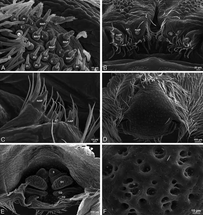
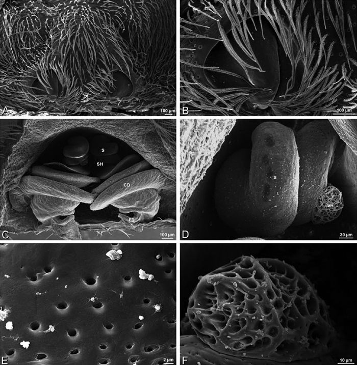
A–F Dresserus sp., scanning electron micrographs. A, C female from Mazumbai, Tanzania (CASENT 9025747, CAS) D, F female from Klein Kariba, South Africa (CASENT 9025745, CAS) A detail of spigots on right ALS B detail of spigots on anterior part of PMS C detail of spigots on anterior part of right PMS D epigynum, ventral view E vulva, dorsal view F detail of pores on right spermathecal head. AC aciniform gland spigot CD copulatory duct CY cylindrical gland spigot FD fertilization duct MAP major ampullate gland spigot mAP minor ampullate gland spigot PI piriform gland spigot S spermatheca SH spermathecal head t tartipore.
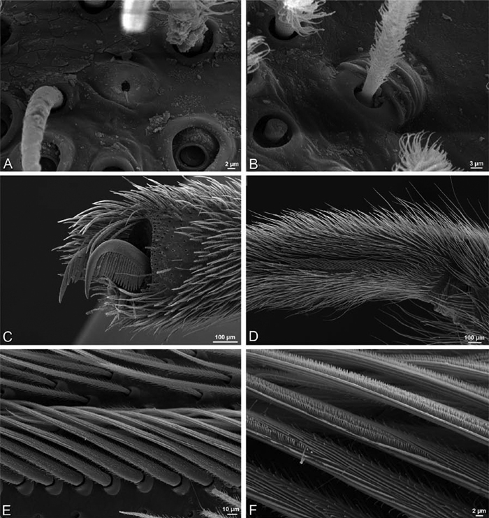
A–F Dresserus sp., female from Klein Kariba, South Africa (CASENT 9025745, CAS), scanning electron micrographs of legs A tarsal organ, left leg I B trichobothrium, left leg I C tarsal claw, left leg I setae removed D left metatarsus IV, retrolateral view, showing calamistrum E detail of calimistrum F detail of teeth on calimistrum setae.
A–F Dresserus sp., male from Mazumbai, Tanzania (CASENT 9025747, CAS), scanning electron micrographs of spinnerets and epiandrous region. A overview of spinnerets B right ALS C PMS D left PLS E modified spigot on right PLS F epiandrous region. AC aciniform gland spigot ALS anterior lateral spinneret MAP major ampullate gland spigot mAP minor ampullate gland spigot MS modified spigot n nubbin PI piriform gland spigot PLS posterior lateral spinneret PMS posterior median spinneret t tartipore.
A–J Eresus walckenaeri. A–D, I–J male from Kresna, Bulgaria (MR) E–H female from 5 km south of Monemvasia, Lakonia, Greece (ZMUC 00012903, ZMUC) A–D habitus of male, photomicrographs E–H habitus of female, photomicrographs I, J illustrations of left male palp A, E dorsal view B, F ventral view C, G anterior view D, H lateral view I prolateral view J retrolateral view. C conductor E embolus ST subtegulum T tegulum.
A–F Eresus walckenaeri from Kresna, Bulgaria (MR), scanning electron micrographs of right male palp, images reversed to appear as left palp. A prolateral view B retrolateral view C detail of embolic division, prolateral view D detail of embolic division, retrolateral view E apex of cymbium, ventral view F detail of embolic division, apical view. C conductor E embolus ST subtegulum T tegulum.
A–F Eresus walckenaeri female from 5 km south of Monemvasia, Lakonia, Greece (ZMUC 00012903), scanning electron micrographs. A prosoma, anterior view B epigynum C tarsal organ, left leg I. ventral view D vulva, dorsal view E left spermatheca F left spermathecal head. S spermatheca SH spermathecal head.
A–J Eresus kollari. A–D, I, J male from Prague, Czechia (MR007, MR) E–H female from res. Srbsko, Czechia (MR016, MR) A–D habitus of male, photomicrographs E–H habitus of female, photomicrographs I, J illustrations of left male palp A, E dorsal view B, F ventral view C, G anterior view D, H lateral view I prolateral view J retrolateral view, arrow indicates notch in conductor. C conductor E embolus T tegulum.
A–F Eresus kollari from Remete Mountain, Hungary (CASENT 9037134, CAS), scanning electron micrographs of left male palp. A prolateral view B retrolateral view C ventral view D palpal bulb, retrolateral view E palpal bulb, retrodorsal view F apical view. C conductor E embolus ST subtegulum T tegulum.
A–F Eresus spp., scanning electron micrographs. A–D Eresus sandaliatus female from SE of Silkeborg, Denmark (CASENT 9039243, CAS) E Eresus kollari female from Srbsko, Czechia (MR016, MR) F Eresus kollari male from Prague, Czechia (MR007, MR) A epigynum, ventral view B vulva, dorsal view C left spermathecal head D detail, left spermatheca E trichobothria, left tibia I F epiandrous region. ML median lobe S spermatheca SH spermathecal head.
A–F Eresus kollari from Srbsko, Czechia (MR016, MR), scanning electron micrographs of female. A prosoma, anterior view B left cheliceral boss C detail of carapace texture D tarsal organ, left leg I E calamistrum, left metatarsus IV F detail, calamistrum seta, left metatarsus IV.
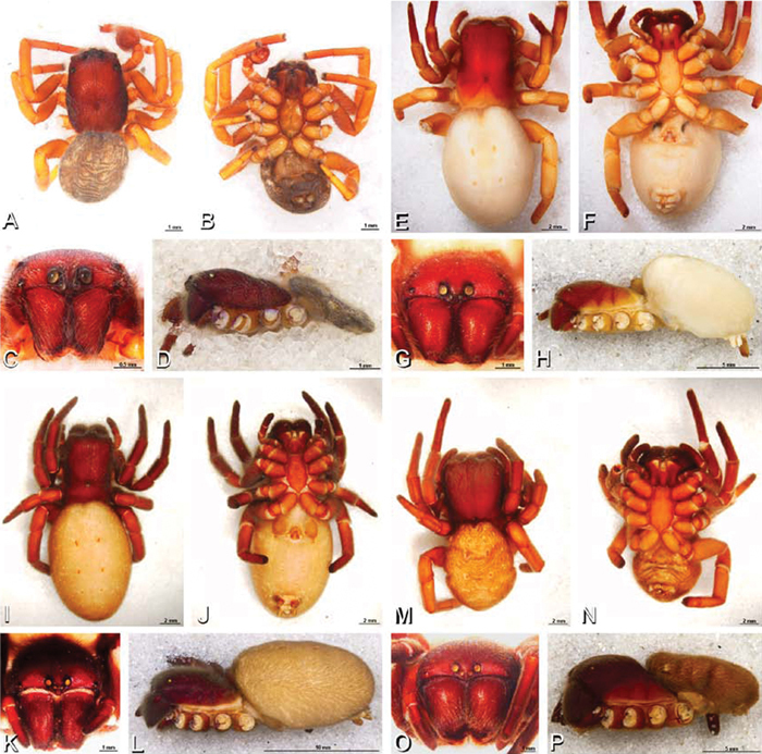
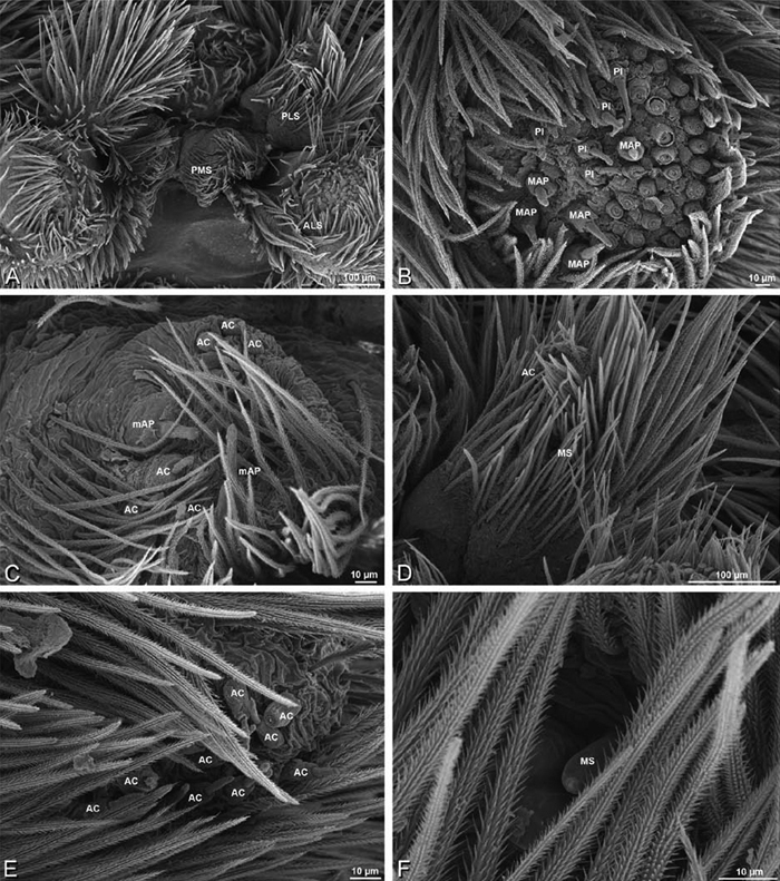
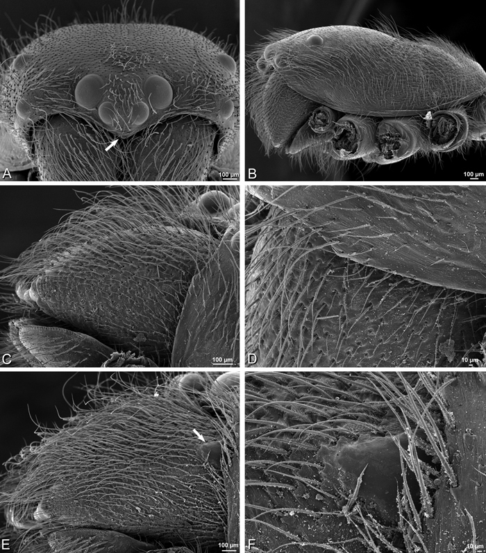
A–P Gandanameno sp., habitus, photomicrographs. A–D male from Harare, Zimbabwe (AcAT 2005/123, NCA), images reversed E–H female from Hanover, South Africa (SAM-ENW-B006896/9958, SAM) I–L female from Iringa, Tanzania (ZMUC 19970530, ZMUC) M–P female from Eierfontein, South Africa (SAM-12823, SAM) A, E, I, M dorsal view B, F, J, N ventral view C, G, K, O anterior view D, H, L, P lateral view.
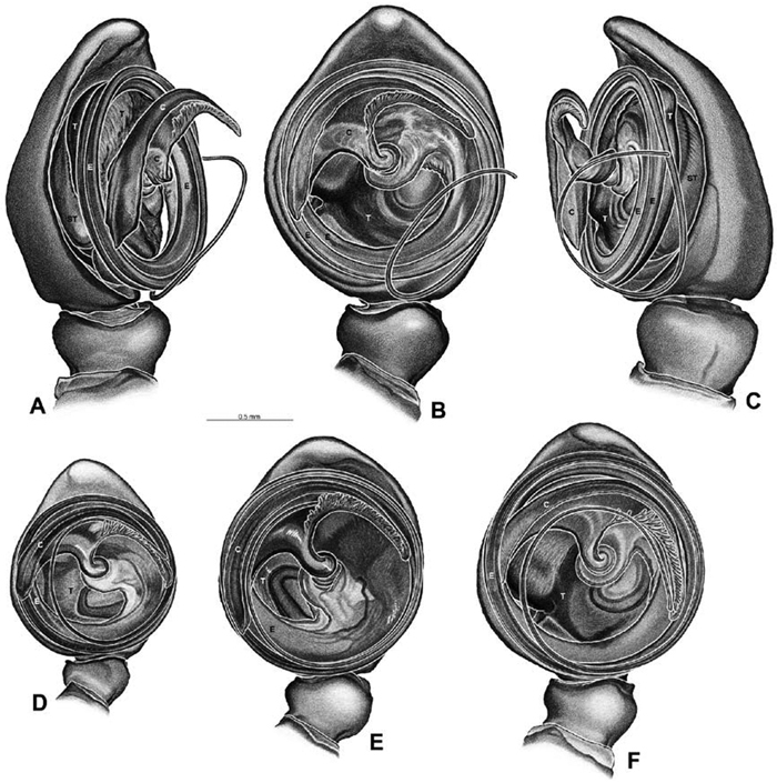
A–F Gandanameno sp., illustrations of left male palp. A–C from Naauwpoort, North West Province, South Africa (SAM 1600, SAM) D from Van Riebeeck Park, Western Cape, South Africa (CASENT 9023763, CAS) E from Graaff-Reinet, Eastern Cape, South Africa (SAM 12571, SAM) F from Hanover, South Africa (SAM 9465, SAM) A obliquely retrolateral view B, D–F ventral view C obliquely prolateral view. All images at the same scale. C conductor E embolus ST subtegulum T tegulum.
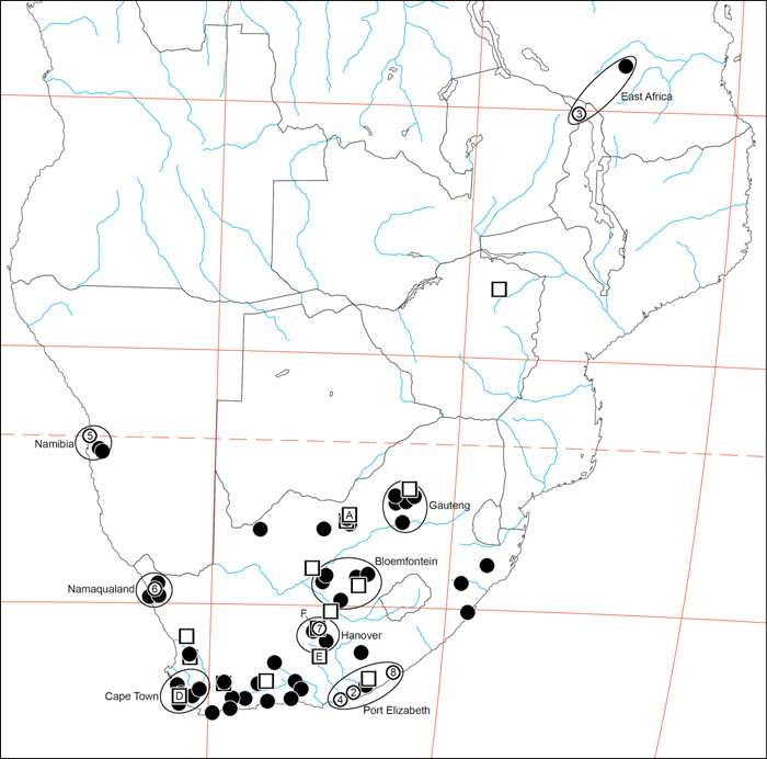
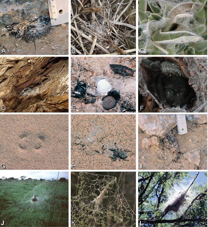
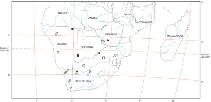
Distribution of Gandanameno. Type localities are numbered circles, males are squares (if with letters, these refer to illustrations in Fig. 48), non-type females are filled circles. Type localities: circle 2 Eresus bubo L. Koch, 1865; circle 3 Eresus inornatus Pocock, 1898; circle 4 Eresus spenceri Pocock, 1900; circle 5 Eresus echinatus Purcell, 1908; circle 6 Eresus namaquensis Purcell, 1908; circle 7 Eresus depressus Tucker, 1920; circle 8 Eresus purcelli Tucker, 1920; type locality of Eresus fumosus C. L. Koch, 1837 is reported simply as “Afrika" and no type specimen is known (Lehtinen 1967: 235). Localities of males illustrated in Fig. 48: square A Fig. 48A–C square D Fig. 48D square E Fig. 48E square F Fig. 48F. Ellipsoids indicate regions for size chart Fig. 50, region names are for convenience only.
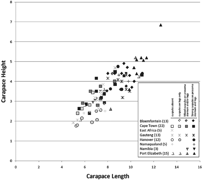
Carapace height plotted against carapace length for adult female Gandanameno specimens from eight regions: Bloemfontein, Cape Town, Gauteng, Hanover, Namaqualand, Namibia, and Port Elizabeth. Regions circumscribed in Fig. 49; sample size given in parentheses. Symbol shape indicates region while symbol darkness indicates presence and strength of cuspules. Specimens were scored as having cuspules absent, having medium to strong cuspules only on the legs, having a mixture of medium and strong cuspules on the prosoma, sternum, and/or legs, and having exclusively strong cuspules on the prosoma, sternum, and legs. As reflected in the legend, not all degrees of spinulation were observed in all regions.
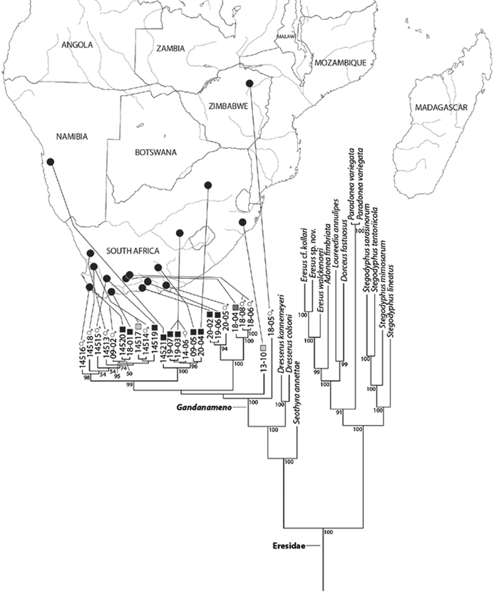
Bayesian phylogenetic tree of the spider family Eresidae based on mixed model analysis (eight data partitions, manually adjusted alignment; see Miller et al. 2010a); outgroups not shown, see Fig. S1. For the genus Gandanameno, DNA specimen codes are substituted for taxonomic name and specimens are linked to their collection locality in southern Africa. Male specimens indicated by male symbol, female specimens indicated either by a female symbol or a square, the darkness of which indicates the strength and presence of cuspules, scored as in Fig. 50. Branches drawn proportional to change. Numbers at nodes are percent posterior probabilities of 50 or greater.
A–F Gandanameno sp., femur, left leg I of female, retrolateral view, scanning electron micrographs. A, C, E overview B, D, F detail of setae A, B from Iringa, Tanzania (ZMUC 19970530, ZMUC) C, D from Hanover, South Africa (SAM-ENW-B006896/9958, SAM) E, F from Eierfontein, Eastern Cape, South Africa (SAM-12823, SAM).
A–F Gandanameno sp., prosoma and coxae of female, scanning electron micrographs. A, C, E detail of setae on prosoma B, F right coxae I and II D left coxae I and II, image reversed to appear as right coxae A, B from Iringa, Tanzania (ZMUC 19970530, ZMUC) C, D from Hanover, South Africa (SAM-ENW-B006896/9958, SAM) E, F from Eierfontein, Eastern Cape, South Africa (SAM-12823, SAM).
A–F Gandanameno sp., sternum of female, scanning electron micrographs A, C, E overview of sternum B, D, F detail of setae on sternum A, B from Iringa, Tanzania (ZMUC 19970530, ZMUC) C, D from Hanover, South Africa (SAM-ENW-B006896/9958, SAM) E, F from Eierfontein, South Africa (SAM-12823, SAM).
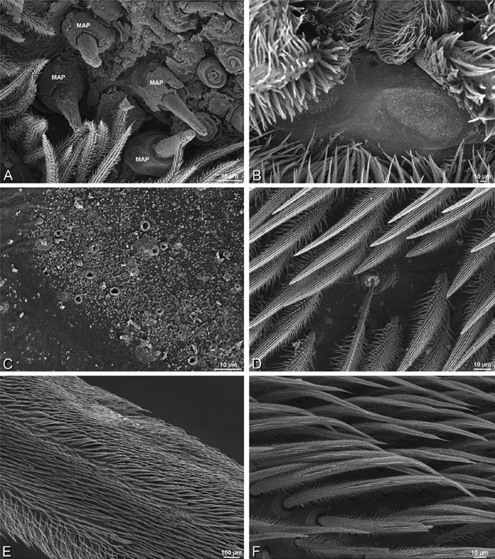
A–F Gandanameno sp. from Harare, Zimbabwe (AcAT 2005/123, NCA), scanning electron micrographs, right male palp, images reversed to appear as left palp. A prolateral view B retrolateral view C ventral view D apical view E detail of distal tip of conductor F palpal tibia, dorsal view. C conductor E embolus ST subtegulum T tegulum.
A–F Gandanameno sp., scanning electron micrographs of prosoma and chelicerae. A–D male from Harare, Zimbabwe (AcAT 2005/123, NCA) E, F male from Hanover, South Africa (SAM 9465, SAM) A prosoma, anterior view, arrow indicates clypeal hood B prosoma, lateral view C, E left chelicerae, lateral view, arrow in E indicates cheliceral boss D detail of left chelicerae showing absence of cheliceral boss F detail of left chelicerae showing cheliceral boss.
A–F Gandanameno sp. from Iringa, Tanzania (ZMUC 19970530, ZMUC), scanning electron micrographs of female spinnerets. A overview B right ALS C right PMS D left PLS E cribellum F cribellar spigots. AC aciniform gland spigot ALS anterior lateral spinneret CR cribellum CY cylindrical gland spigot MAP major ampullate gland spigot mAP minor ampullate gland spigot MS modified spigot n nubbin PI piriform gland spigot PLS posterior lateral spinneret PMS posterior median spinneret t tartipore.
A–F Gandanameno sp., scanning electron micrographs. A–E female from Iringa, Tanzania (ZMUC 19970530, ZMUC) F female from Hanover, South Africa (SAM-ENW-B006896/9958) A, B prosoma C–F details of spinneret spigots A anterior view, arrow indicates clypeal hood B left cheliceral boss C detail of modified spigots on right female PLS D detail of spigots on anterior part of right female PMS E detail of cylindrical gland spigots on posterior part of left female PMS F right PMS. AC aciniform gland spigot CY cylindrical gland spigot MAP major ampullate gland spigot mAP minor ampullate gland spigot MS modified spigot n nubbin PI piriform gland spigot t tartipore.
A–F Scanning electron micrographs of epigynum and vulva of Gandanameno sp. A, B from Iringa, Tanzania (ZMUC 19970530, ZMUC) C–F from Kommetjie, Cape Town, South Africa (CASENT 9039241, CAS) A epigynum, ventral view B detail of right copulatory opening, ventral view C cleared vulva, dorsal view D detail of right spermatheca and spermathecal head E detail, right spermatheca F detail, right spermathecal head. CD copulatory duct FD fertilization duct S spermatheca SH spermathecal head.
A–F Gandanameno sp. from Hanover, South Africa (SAM 9465, SAM), scanning electron micrographs of male spinnerets. A overview B left ALS C right PMS D left PLS E vestigial cribellum F detail of vestigial cribellum. AC aciniform gland spigot ALS anterior lateral spinneret MAP major ampullate gland spigot mAP minor ampullate gland spigot MS modified spigot PI piriform gland spigot PLS posterior lateral spinneret PMS posterior median spinneret n nubbin t tartipore.
A–F Gandanameno sp. from Hanover, South Africa (SAM 9465, SAM), scanning electron micrographs of male spinnerets. A detail of spigots on left ALS B left PLS C detail of spigots on left PLS D detail of spigots on left PMS E epiandrous region F detail of epiandrous gland spigots. AC aciniform gland spigot MAP major ampullate gland spigot mAP minor ampullate gland spigot MS modified spigot PI piriform gland spigot.
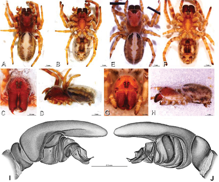
A–J Loureedia annulipes. A, B, D I, J male from Haluqim Ridge, Israel (MR008, HUJ) C male from Nitzanna village, Israel (MR018, HUJ) E, F, H female from Wadi Mashash, Israel (MR019, MR) G female from Haluquim, Israel (PET03, MR) A–D habitus of male, photomicrographs E–H habitus of female, photomicrographs I–J illustrations of left male palp A, E dorsal view B, F ventral view C, G anterior view D, H lateral view I prolateral view J retrolateral view. C conductor E embolus T tegulum.
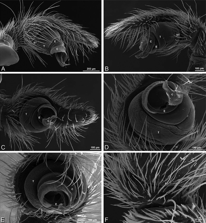
A–F Loureedia annulipes from Haluqim Ridge, Israel (MR008, HUJ), scanning electron micrographs of right male palp, images reversed to appear as left palp. A prolateral view B retrolateral view C conductor, prolateral view D ventral view E conductor, ventral view F apical view. C conductor E embolus ST subtegulum T tegulum.
A–F Loureedia annulipes, scanning electron micrographs of female from from Wadi Mashash, Negev, Israel (MR019, MR), images reversed. A prosoma, anterior view B chelicera C cheliceral boss D sternum, ventral view.
A–F Loureedia annulipes, scanning electron micrographs. A–E vulva of female from Wadi Mashash, Negev, Israel (MR019, MR) F male from Haluqim Ridge, Israel (MR008, HUJ) A epigynum, ventral view B cleared vulva, dorsal view C, D detail, left spermathecal head E detail, right spermatheca. F epiandrous region. AL anterior lobe on atrium ML median lobe S spermatheca SH spermathecal head.
A–F Loureedia annulipes, female from Wadi Mashash, Negev, Israel (MR019, MR), scanning electron micrographs of spinnerets. A overview B right ALS C left and right PMS D right PLS E cribellum. F cribellar spigots. Unlabeled spigots in C thought to be a mixture of aciniform gland spigots and cylindrical gland spigots. AC aciniform gland spigot ALS anterior lateral spinneret MAP major ampullate gland spigot mAP minor ampullate gland spigot MS modified spigot PI piriform gland spigot PLS posterior lateral spinneret PMS posterior median spinneret.
A–F Loureedia annulipes, female from Wadi Mashash, Negev, Israel (MR019, MR), scanning electron micrographs of spinnerets. A detail of spigots on right ALS B, C detail of spigots on left PMS D modified spigot and flanking aciniform gland spigots on left PLS E calamistrum, left metatarsus IV F detail, calamistrum seta, left metatarsus IV. Spigots in B thought to be a mixture of aciniform gland spigots and cylindrical gland spigots; unlabeled spigot in C thought to be either an aciniform gland spigot or a cylindrical gland spigot. AC aciniform gland spigot MAP major ampullate gland spigot mAP minor ampullate gland spigot MS modified spigot PI piriform gland spigot.
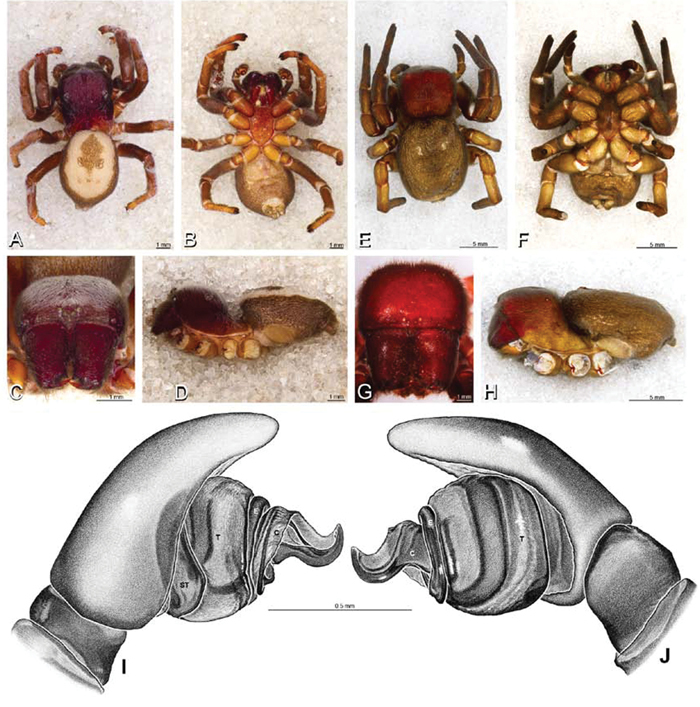
A–J males of Paradonea striatipes and Paradonea splendens. A–C, G, H Paradonea striatipes, male from Otjivasandu, Namibia (NMN) D–F, I, J Paradonea splendens, male from Sunnyside, South Africa (C1076, SAM) A–F habitus of male, photomicrographs G–J illustrations of left male palp A, D dorsal view B, E ventral view C, F anterior view G, I prolateral view H, J retrolateral view. C conductor E embolus ST subtegulum T tegulum.
A–H Paradonea variegata. A–C, G–H male from Breekkierie Dunes, Northern Cape, South Africa (C1062, SAM) D–F female from Steinkopf, South Africa (ZMB 26964, ZMHB) A–C habitus of male, photomicrographs D–F habitus of female photomicrographs G–H illustrations of left male palp A, D dorsal view B, E ventral view C, F anterior view G prolateral view H retrolateral view. C conductor ST subtegulum T tegulum.
A–I males of Paradonea parva and Paradonea presleyi sp. n., habitus, photomicrographs. A–F Paradonea parva A–C male holotype from junction of Marico and Crocodile Rivers, South Africa (B3701, SAM) D–F male from 4 km N of Hopetown, Northern Cape, South Africa (AcAT 97/988, NCA). G–I male holotype of Paradonea presleyi sp. n. from Falcon College, Zimbabwe (CASENT 9039236, CAS) A, D, G dorsal view B, E, H ventral view C, F, I anterior view.
Distribution of Paradonea species. Circles, Paradonea striatipes; squares, Paradonea splendens; inverted triangles, Paradonea variegata; diamonds, Paradonea parva; triangles, Paradonea presleyi sp. n. Primary type localities with all black symbols, those for other localities with white center.
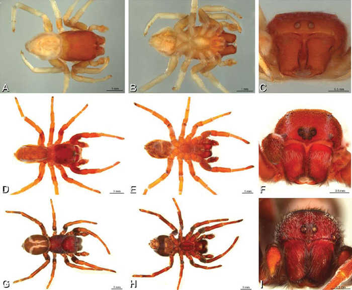
A–J Seothyra henscheli. A–D, I–J male from Gobabeb Station, Namibia (SMN 40828, NMN) E–H female from Kuiseb River, Gobabeb, Namibia (SMN 46627, NMN) A–D habitus of male, photomicrographs E–H habitus of female, photomicrographs I, J illustrations of left male palp A, E dorsal view B, F ventral view C, G anterior view D, H lateral view I prolateral view J retrolateral view. C conductor E embolus ST subtegulum T tegulum.
A–F Seothyra henscheli from Gobabeb Station, Namibia (SMN 40828, NMN), scanning electron micrographs of right male palp, images reversed to appear as left palp. A prolateral view B retrolateral view C detail of embolic division, prolateral view D detail of modified setae on retrolateral side of cymbium. E ventral view F apical view. C conductor E embolus ST subtegulum T tegulum.
A–D Seothyra henscheli from Gobabeb Station, Namibia (SMN 40828, NMN), scanning electron micrographs of male legI spinnerets, and epiandrous region. A femurI retrolateral view B detail of modified spigot on left male PLS C epiandrous region D detail of epiandrous gland spigots. MS modified spigot.
A–F Seothyra henscheli from Kuiseb River, Gobabeb, Namibia (SMN 46627, NMN), scanning electron micrographs of female prosoma and spinnerets. A prosoma, anterior view, arrow indicates clypeal hood B left chelicerae, ectal view C detail of setae on prosoma D sternum, ventral view E detail of piriform gland spigots on left female ALS F detail of modified spigot on right female PLS. MS modified spigot PI piriform gland spigot.
A–F Seothyra henscheli from Sout Rivier, Namibia (CASENT 9039242, CAS), scanning electron micrographs of epigynum A epigynum, ventral view B vulva, dorsal view C, D spermathecal head. ML median lobe S spermatheca SH spermathecal head.
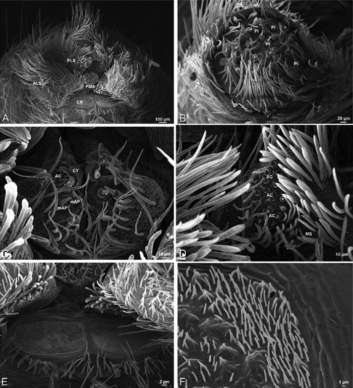
Figure 77. A–F Seothyra henscheli from Kuiseb River, Gobabeb, Namibia (SMN 46627, NMN), scanning electron micrographs of female spinnerets. A overview B left ALS C left and right PMS D right PLS E cribellum F cribellar spigots. AC aciniform gland spigot ALS anterior lateral spinneret CR cribellum CY cylindrical gland spigot mAP minor ampullate gland spigot MS modified spigot PI piriform gland spigot PLS posterior lateral spinneret PMS posterior median spinneret.
A–F Seothyra henscheli from Gobabeb Station, Namibia (SMN 40828, NMN), scanning electron micrographs of male spinnerets. A overview B right ALS C left PMS D left PLS E vestigial cribellum F detail of vestigial cribellum. AC aciniform gland spigot ALS anterior lateral spinneret mAP minor ampullate gland spigot MS modified spigot PI piriform gland spigot PLS posterior lateral spinneret PMS posterior median spinneret.
A–J Stegodyphus lineatus. A–D, I, J male from Nengrahar, Afghanistan (MR010, MR) E–H female from Belkis, near Birecor, Turkey (MR015, MR) A–D habitus of male, photomicrographs E–H habitus of female, photomicrographs I, J illustrations of left male palp A, E dorsal view B, F ventral view C, G anterior view D, H lateral view I prolateral view J retrolateral view. C conductor E embolus T tegulum.
A–D male Stegodyphus lineatus from Nengrahar, Afghanistan (MR010, MR), scanning electron micrographs. A–D right palp, images reversed to appear as left palp E, F epiandrous region A prolateral view B retrolateral view C apical view D detail, prolateral view E ventral view F detail of epiandrous gland spigots. C conductor E embolus ST subtegulum T tegulum.
A–F Female Stegodyphus lineatus from Belkis, near Birecor, Turkey (MR015, MR), scanning electron micrographs of prosoma and chelicerae. A anterior view, arrow indicates clypeal hood B left lateral view C eye region, dorsal view D detail, prosoma cuticle E left chelicera F left cheliceral boss.
A–F female Stegodyphus lineatus from Belkis, near Birecor, Turkey (MR015, MR), scanning electron micrographs of epigynum, vulva, and calamistrum. A epigynum, ventral view B vulva, dorsal view C right spermatheca and fertilization duct D right spermathecal head E calamistrum, left metatarsus IV F detail, calamistrum setae. FD fertilization duct ML median lobe; S spermatheca SH spermathecal head.
A–F Stegodyphus lineatus, scanning electron micrographs of spinnerets and trichobothrium. A–D male from Nengrahar, Afghanistan (MR010, MR) E, F female from Belkis, near Birecor, Turkey (MR015, MR). A overview B, E right ALS C left PMS D right PLS F trichobothrium on left metatarsus I. AC aciniform gland spigot ALS anterior lateral spinneret MAP major ampullate gland spigot mAP minor ampullate gland spigot PI piriform gland spigot PLS posterior lateral spinneret PMS posterior median spinneret.
A–J Stegodyphus mimosarum from Forêt d'Analalava, Fianarantsoa, Madagascar (CASENT 9005869, CAS), images reversed. A–D, I, J male E–H female A–D habitus of male, photomicrographs E–H habitus of female, photomicrographs J–K illustrations of left male palp A, E dorsal view B, F ventral view C, G anterior view D, H lateral view I prolateral view J retrolateral view. C conductor E embolus T tegulum.
A–F Stegodyphus mimosarum, scanning electron micrographs of male from Forêt d'Analalava, Fianarantsoa, Madagascar (CASENT 9015950, CAS). A–D right palp, images reversed so as to appear a left palp E, F epiandrous gland spigots A prolateral view B retrolateral view C apical view D ventral view E epiandrous region, ventral view F detail, epiandrous gland spigots. C conductor E embolus T tegulum.
A–D Stegodyphus mimosarum female from Forêt d'Analalava, Fianarantsoa, Madagascar (CASENT 9015950, CAS), scanning electron micrographs of genitalia. A epigynum, ventral view B vulva, dorsal view C spermathecal head D spermatheca and fertilization duct. FD fertilization duct ML median lobe S spermatheca SH spermathecal head.
A–F Stegodyphus mimosarum female from Forêt d'Analalava, Fianarantsoa, Madagascar (CASENT 9015950, CAS), scanning electron micrographs of spinnerets. A Overview B Left ALS C Left PMS D Right PLS E Left lobe of cribellum F Cribellar spigots. Unlabeled spigots in C thought to be a mixture of aciniform gland spigots and cylindrical gland spigots. AC aciniform gland spigot ALS anterior lateral spinneret CR cribellum MAP major ampullate gland spigot mAP minor ampullate gland spigot MS modified spigot PI piriform gland spigot PLS posterior lateral spinneret PMS posterior median spinneret t tartipore.
A–F Stegodyphus mimosarum male from Forêt d'Analalava, Fianarantsoa, Madagascar (CASENT 9015950, CAS), scanning electron micrographs of spinnerets. A Overview B Right ALS C Left PMS D Right PLS E Detail of modified spigot on right PLS F Vestigial cribellum. AC aciniform gland spigot ALS anterior lateral spinneret CR cribellum MAP major ampullate gland spigot mAP minor ampullate gland spigot MS modified spigot PI piriform gland spigot PLS posterior lateral spinneret PMS posterior median spinneret t tartipore.
A–J Stegodyphus sarasinorum from 7.5 km E PwintPhyu, Magway Division, Myanmar (CASENT 9019370, CAS). A–D, I–J male, images reversed E–H female A–D habitus of male, photomicrographs E–H habitus of female, photomicrographs I, J illustrations of left male palp A, E dorsal view B, F ventral view C, G anterior view D, H lateral view I prolateral view J retrolateral view. C conductor E embolus T tegulum.
A–F Stegodyphus sarasinorum male from 7.5 km E PwintPhyu, Magway Division, Myanmar (CASENT 9019370, CAS), scanning electron micrographs of right palp, images reversed to appear as left palp. A prolateral view B retrolateral view C ventral view D ventral-apical view E apical view F palpal tibia, dorsal view. C conductor E embolus ST subtegulum T tegulum.
A–F Stegodyphus sarasinorum female from 7.5 km E PwintPhyu, Magway Division, Myanmar (CASENT 9019370, CAS), scanning electron micrographs of prosoma and chelicerae. A Prosoma, anterior view, left chelicera removed, arrow indicates clypeal hood B prosoma, dorsal view C sternum, ventral view D left chelicerae, ectal view E left cheliceral boss F distal part of left chelicera showing fang and teeth.
A–F, Stegodyphus sarasinorum, scanning electron micrographs of female from 7.5 km E PwintPhyu, Magway Division, Myanmar (CASENT 9019370, CAS). A tarsal organ indicated by arrow, left leg I B trichobothrium, left metatarsus I C tarsal claws, left legI retrolateral view D calamistrum, left metatarsus IV E detail of calamistrum setae F left female palp, retrolateral view.
A–F, Stegodyphus sarasinorum from 7.5 km E PwintPhyu, Magway Division, Myanmar (CASENT 9019370, CAS), scanning electron micrographs. A–E female F male A epigynum, ventral view B vulva, dorsal view C vulva, dorsolateral view D detail of spermatheca E detail of spermathecal head F epiandrous gland spigots. FD fertilization duct ML median lobe S spermatheca SH spermathecal head.
A–F Stegodyphus sarasinorum, scanning electron micrographs of spinnerets of female from 7.5 km E PwintPhyu, Magway Division, Myanmar (CASENT 9019370, CAS). A overview B right ALS C left PMS D right PLS E cribellum F cribellar spigots. Unlabeled spigots in C thought to be a mixture of aciniform gland spigots and cylindrical gland spigots. AC aciniform gland spigot ALS anterior lateral spinneret CR cribellum MAP major ampullate gland spigot mAP minor ampullate gland spigot MS modified spigot n nubbin PI piriform gland spigot PLS posterior lateral spinneret PMS posterior median spinneret t tartipore.
A–F Stegodyphus sarasinorum, spinnerets of male from 7.5 km E PwintPhyu, Magway Division, Myanmar (CASENT 9019370, CAS). A overview B right ALS C right PMS D right PLS E detail of modified spigot on right PLS F vestigial cribellum. AC aciniform gland spigot ALS anterior lateral spinneret MAP major ampullate gland spigot mAP minor ampullate gland spigot MS modified spigot PI piriform gland spigot PLS posterior lateral spinneret PMS posterior median spinneret t tartipore.