Research Article |
|
Corresponding author: Somsak Panha ( somsak.pan@chula.ac.th ) Academic editor: Fredric Govedich
© 2014 Jaruwan Tubtimon, Ekgachai Jeratthitikul, Chirasak Sutcharit, Bangon Kongim, Somsak Panha.
This is an open access article distributed under the terms of the Creative Commons Attribution License (CC BY 4.0), which permits unrestricted use, distribution, and reproduction in any medium, provided the original author and source are credited.
Citation:
Tubtimon J, Jeratthitikul E, Sutcharit C, Kongim B, Panha S (2014) Systematics of the freshwater leech genus Hirudinaria Whitman, 1886 (Arhynchobdellida, Hirudinidae) from northeastern Thailand. ZooKeys 452: 15-33. https://doi.org/10.3897/zookeys.452.7528
|
Abstract
In total, 435 specimens of the Southeast Asian freshwater leech species within the Hirudinidae family were collected from 17 locations of various types of aquatic habitats in northeastern Thailand. They were all morphologically placed within the genus Hirudinaria Whitman, 1886 and there were three distinct species: the common Hirudinaria manillensis, 78.2% of all collected specimens and at all 17 locations, Hirudinaria javanica at 20.3% of collected samples and from five locations and a rarer unidentified morphospecies (Hirudinaria sp.) with six samples from only two locations. The karyotypes of these three species were examined across their range in this study area for 38, 11 and 6 adult specimens of Hirudinaria manillensis, Hirudinaria javanica and Hirudinaria sp., respectively. This revealed different chromosome numbers among all three species, with Hirudinaria javanica having n = 13, 2n = 26, Hirudinaria manillensis lacked one small chromosome pair with n = 12, 2n = 24, and the unknown Hirudinaria sp. differed from any known Hirudinaria karyotypes in exhibiting a higher chromosome number (n = 14, 2n = 28) and a gradual change in size from large to small chromosomes. This suggests that the unknown Hirudinaria sp. is a new biological species. However, phylogenetic analysis based upon a 658 bp fragment of the cytochrome oxidase subunit I gene placed this unknown morphospecies within the Hirudinaria manillensis clade, perhaps then suggesting a recent sympatric speciation, although this requires further confirmation. Regardless, the chromosomes of all three species were asymmetric, most with telocentric elements. A distinct bi-armed chromosome marker was present on the first chromosome pair in Hirudinaria javanica, whilst it was on pairs 1, 2, 3 and 5 in Hirudinaria manillensis, and on pairs 3 and 5 for the unknown Hirudinaria sp.
Keywords
Freshwater leeches, Hirudinea , karyotypes, morphology, COI, sanguivorous
Introduction
The family Hirudinidae (Arhynchobdellida, Hirudiniformes) is comprised of mainly blood-sucking (sanguivorous) freshwater leeches, or medicinal leeches, although four terrestrial species are known. It includes approximately 60 hirudinids ranging across all continents, except for Antarctica, and from temperate to tropical regions (
The genus Hirudinaria Whitman, 1886 consists of only three known species Hirudinaria javanica (Wahlberg, 1856), Hirudinaria manillensis (Lesson, 1842), and Hirudinaria bpling that are widely distributed over tropical South and Southeast Asia, being recorded from within Peninsular Malaysia, Thailand, Indo-China, Indonesia, Philippines, China, Myanmar, Bangladesh, India and Sri Lanka (
In this study, we examined the karyotypes of 38, 11 and 6 specimens of the three species (H. manillensis, H. javanica, and a third distinct and different morphospecies, Hirudinaria sp.) collected from across 17 locations in northeastern Thailand, representing 13.4%, 12% and 100% of the collected samples, respectively. Their systematic implications are then discussed in comparison with other previously reported hirudinid karyotypes. The phylogenetic analysis, based upon a 658 bp fragment of the cytochrome oxidase subunit I gene, was also conducted to clarify the systematics of all collected morphospecies.
Materials and methods
Locality, co-ordination and sample size for all collected species are given in Table
Freshwater leeches were collected from 17 localities in northeastern Thailand (Fig.

Locality, co-ordination and sample size of each species used in the present study. Locality numbers refer to the localities shown in Figure
| No. | Locality | Coordinates | Number of specimens examined | ||
|---|---|---|---|---|---|
| Hirudinaria javanica | Hirudinaria manillensis | Hirudinaria sp. | |||
| 1 | Ban Donsala, Na Wa, Nakhon Phanom | 17°34'27.22"N, 104°7'18.64"E | 44 | 82 | 5 |
| 2 | Ban Majang, Na Wa, Nakhon Phanom | 17°36'53.4"N, 104°8'21.9"E | - | 51 | 1 |
| 3 | Ban Nongwang, Tao Ngoi, Sakon Nakhon | 17°45'41.26"N, 103°44'42.00"E | 9 | 4 | - |
| 4 | Phang Khon, Sakon Nakhon | 17°22'29.02"N, 103°40'26.81"E | - | 2 | - |
| 5 | Mueang, Sakon Nakhon | 17°10'52.69"N, 104°7'50.94"E | - | 2 | - |
| 6 | Phu Phan, Sakon Nakhon | 16°54'14.64"N, 103°54'7.50"E | - | 6 | - |
| 7 | Ban Janpen, Tao Ngoi, Sakon Nakhon | 16°55'32.59"N, 104°10'9.31"E | 16 | 1 | - |
| 8 | Ban Nonghai, Khamcha-i, Mukdahan | 16°34'53.92"N, 104°29'29.00"E | 13 | 13 | - |
| 9 | Khong Chai, Kalasin | 16°15'44.76"N, 103°27'22.91"E | - | 28 | - |
| 10 | Ban Thatoom, Mueang, Mahasarakham | 16°10'48.40"N, 103°26'59.30"E | - | 4 | - |
| 11 | Huai E-pong, Phu Wiang, Khon Kaen | 16°43'51.30"N, 102°17'17.00"E | - | 11 | - |
| 12 | Tumbon Bung, Mueang Amnat Charoen | 15°50'21.48"N, 104°27'33.95"E | - | 30 | - |
| 13 | Pa Tio, Yasothon | 15°57'2.81"N, 104°25'12.78"E | - | 3 | - |
| 14 | Khemarat, Ubon Ratchathani | 15°59'11.82"N, 105°8'20.53"E | - | 26 | - |
| 15 | Chaturaphak Phiman, Roi Et | 15°49'59.77"N, 103°31'0.86"E | 1 | 5 | - |
| 16 | Kaset Wisai, Roi Et | 15°39'13.70"N, 103°35'58.39"E | - | 67 | - |
| 17 | Huai Saneng Reservoir, Surin | 14°47'14.70"N, 103°28'34.50"E | - | 11 | - |
Jaws of some specimens were examined by scanning electron microscopy (SEM). The dried specimens were sputter coated with 35 nm of gold/palladium before being examined using a LEO/Zeiss DSM982 Geminifield emission scanning electron microscope located in the Scientific and Technological Research Equipment Centre, Chulalongkorn University.
Chromosome preparations were made from the testisac using hypotonic, fixation and air-drying techniques modified from
For the molecular analysis, the total genomic DNA was extracted from a part of the wall-body muscle to avoid contamination from the host DNA, following the standard protocol of the DNeasy Blood & Tissue Kit (Qiagen Inc., Valencia, CA, USA). A fragment of the mitochondrial cytochrome oxidase subunit I (COI) gene was amplified using the primers LCO1490 (5’-GGT CAA CAA ATC ATA AAG ATA TTG G-3’) and HCO2198 (5’-TAA ACT TCA GGG TGA CCA AAA AAT CA-3’), which is the region used in animal DNA barcoding (
Sequence alignment and editing were performed using MEGA 6.06 (
Nucleotide sequences obtained in this study have been deposited in the GenBank database under the GenBank ID: KJ551848–KJ551855.
Results
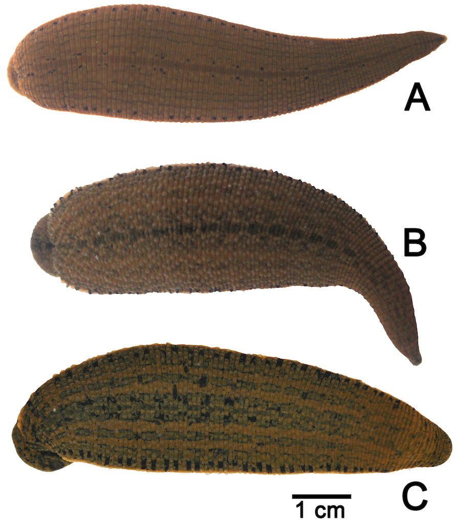
All 435 examined specimens in this study were assigned as belonging to the genus Hirudinaria by the following distinct characters; male pore and female pore separated by 5–7 annuli, sensillae large and elongated, salivary papillae present, and without vaginal stalk. From these identified characters, the specimens were determined to be three species: as Hirudinaria javanica, Hirudinaria manillensis, and an unidentified morphotype, Hirudinaria sp. (Figs

Systematics
Family Hirudinidae Whitman, 1886
Subfamily Hirudininae
Hirudinaria
Hirudinaria Whitman, 1886: 373.
Type species
Sanguisuga javanica Wahlberg, 1856, by original designation.
Hirudinaria javanica
Sanguisuga javanica Wahlberg, 1856: 233. Type locality: Samarang, Java [Semarang, Central Java, Indonesia].
Hirudinaria javanica –
Limnatis (Poecilobdella) javanica –
Limnatis javanica –
Hirudinaria javanica –
Material examined
Ban Donsala, Na Wa, Nakhon Phanom: CUMZ 3402 (17 specimens), 3404 (18 specimens; Figs

Illustrations of the reproductive system of A Hirudinaria javanica CUMZ 3404 from Nakhon Phanom, B Hirudinaria manillensis CUMZ 3403 from Nakhon Phanom, and C Hirudinaria sp. CUMZ 3405 from Nakhon Phanom. Abbreviations are: ag = albumin gland, at = atrium, cod = common oviduct, eb = ejaculatory bulb, ep = epididymis, g = ganglion, o = ovary, ps = penis sheath, vas = vas deferens, vc = vagina sac, vd = vagina duct.


Description
In preserved specimens body length 41–184 mm, width 5–16 mm. In live specimens, dorsal side olive green, dark green or yellow brown. Middle dorsal line distinct, black, continuous, parallel with two series of black spots on both sides, two faint black stripes present on each side. Body margin yellow with one ordered series of black spots. Ventral side green without marker. Jaw trignathous, approximately 134 teeth. Number of salivary papillae, both small and large, is 43 glands (Fig.
Hirudinaria manillensis
Hirudo manillensis Lesson, 1842: 8. Type locality: Philippine Islands.
Hirudo sanguisorba Tennent, 1859: 305. Type locality: Ceylon.
Hirudo multistriata Schmarda, 1861: 3, Taf. 16, fig. 141. Type locality: Ceylon [Sri Lanka].
Hirudo luzoniae Kinberg, 1866: 356. Type locality: Manila [Philippines].
Hirudo maculosa Grube, 1868: 39–40, Taf. 4, fig. 6. Type locality: Singapore.
Hirudo maculata Baird, 1869: 315. Type locality: Siam [Thailand].
Limnatis (Poecilobdella) granulosa Blanchard, 1893: 28. Type locality: Java, Indonesia
Limnatis granulosa –
Hirudo boyntoni Wharton, 1913: 369–371. Type locality: Philippines Islands.
Limnatis maculosa –
Limnatis (Poecilobdella) manillensis –
Hirudinaria manillensis –
Material examined
Ban Donsala, Na Wa, Nakhon Phanom: CUMZ 3401 (21 specimens), 3403 (4 specimens; Figs
Description
In preserved specimens, body length 27–248 mm, width 3–30 mm. In live specimens, dorsal side dark green or brown. Middle dorsal line distinct, black, incontinuous, with two faint black stripes on each side. Body margin yellow with disrupted black spots. Ventral side brown without marker. Jaw trignathous, approximately 148 teeth. Number of salivary papillae, both small and large sizes, is 30 glands (Fig.
Hirudinaria sp.
Material examined
Ban Donsala, Na Wa, Nakhon Phanom: CUMZ 3405 (1 specimen; Figs
Description
In preserved specimens body length 107–140 mm, width 11–16 mm. In live specimens, dorsal side dark green, brown and dark brown. Middle dorsal line not present. Two brown stripes present each side of mid-dorsal region. Body margin yellow or orange with one ordered series of short black lines. Ventral side brown or dark brown without marker. Jaw trignathous, approximately 167 teeth. Number of salivary papillae, both small and large sizes, is 25 glands (Fig.
Karyotype results
The chromosomes were typically indistinct because of their small size. Nevertheless, all cleared metaphase arrangements could be observed and the spermatogonial meiotic and mitotic chromosome numbers could be confirmed for all the examined species (Fig.
Comparative morphological characters among Hirudinaria species in this study.
| Characters | Hirudinaria bpling | Hirudinaria javanica | Hirudinaria manillensis | Hirudinaria sp. |
|---|---|---|---|---|
| Color | dark brown | dark green | dark brown/brown | dark green/brown |
| Distance (annuli) between male & female pores | 5 | 7 | 5 | 5 |
| Position of male and female organs | XI-XII | XI-XIII | XI-XII | XI-XII |
| Atrium | bulbous | short | long | relative long |
| Pairs of testisacs | - | 12 | 11 | 11 |
| Common oviduct | short | short | short | long |
| Vagina caecum | wide, long | small, ovate | small, ovate | large, elongate |
| References |
|
This study | This study | This study |
| Species | Locality no. |
No. |
Haploid (n) | Diploid (2n) | Reference |
|---|---|---|---|---|---|
| Hirudo medicinalis | Kharkiv, Ukraine | 5 | 14 | 28 |
|
| Hirudo verbana | Odesa and Kharkiv, Ukraine | 6 | 13 | 26 |
|
| Hirudo orientalis | Lake Taskul, Kazakhstan | 7 | 12 | 24 |
|
| Hirudinaria javanica | 1 | 4 | 13 | 26 | This study |
| 3 | 1 | 13 | 26 | This study | |
| 7 | 2 | 13 | 26 | This study | |
| 8 | 3 | 13 | 26 | This study | |
| 15 | 1 | 13 | 26 | This study | |
| Hirudinaria manillensis | 1 | 5 | 12 | 24 | This study |
| 2 | 3 | 12 | 24 | This study | |
| 3 | 1 | 12 | 24 | This study | |
| 4 | 1 | 12 | 24 | This study | |
| 5 | 1 | 12 | 24 | This study | |
| 6 | 2 | 12 | 24 | This study | |
| 8 | 2 | 12 | 24 | This study | |
| 9 | 2 | 12 | 24 | This study | |
| 10 | 2 | 12 | 24 | This study | |
| 11 | 2 | 12 | 24 | This study | |
| 12 | 3 | 12 | 24 | This study | |
| 13 | 2 | 12 | 24 | This study | |
| 14 | 4 | 12 | 24 | This study | |
| 16 | 4 | 12 | 24 | This study | |
| 17 | 4 | 12 | 24 | This study | |
| Hirudinaria sp. | 1 | 5 | 14 | 28 | This study |
| 2 | 1 | 14 | 28 | This study |
The karyotypes of all three species were asymmetric, and mostly telocentric, chromosomes. The distinct bi-armed chromosome marker varied among the three species, being found on the first pair in Hirudinaria javanica, on pairs 1, 2, 3 and 5 for Hirudinaria manillensis and on pairs 3 and 5 for Hirudinaria sp.

Phylogenetic analysis
The samples used for phylogenetic analysis and their collection locations are summarized in Table

Phylogenetic relationships of the genus Hirudinaria and their related species, with chromosome number data. Tree topology was obtained from ML analysis based on a 658 bp fragment of the mitochondrial COI gene (DNA barcode region). Nodes with a 0.95 or higher bipartition posterior probability for BI and/or 70% or higher bootstrap value for ML were regarded as sufficiently resolved nodes, and are shown for the major clades (ML/BI). Numbers in parentheses refer to sampling localities in Figure
Taxa examined in the phylogenetic analysis, with collection localities and COI GenBank accession numbers.
| Taxon | Locality no. |
Gen Bank accession nos. |
|---|---|---|
| Hirudinaria javanica (2n = 26) | 1 | KJ551852 |
| 7 | KJ551853, KJ551854 | |
| 8 | KJ551851 | |
| 15 | KJ551855 | |
| Hirudinaria manillensis (2n = 24) | 1 | KJ551850 |
| 5 | KJ551850 | |
| 17 | KJ551850 | |
| Hirudinaria sp. (2n = 28) | 1 | KJ551848 |
| 2 | KJ551849 | |
| Hirudinaria bpling | Phang Nga, Thailand |
JQ846012
|
| Haemopis sanguisuga | Sweden |
AF462021
|
| Hirudo verbana | USA |
GQ368752
|
| Hirudo orientalis | - |
JN104645
|
| Hirudo medicinalis | Sweden |
HQ333518
|
The uncorrected p-distances between the members of the genus Hirudinaria are shown in Table
Average uncorrected p-distance for the 658 bp COI gene sequences of the genus Hirudinaria.
| Speceis | 1 | 2 | 3 | 4 |
|---|---|---|---|---|
| 1. Hirudinaria javanica (2n = 26) | - | |||
| 2. Hirudinaria manillensis (2n = 24) | 0.101 | - | ||
| 3. Hirudinaria sp. (2n = 28) | 0.110 | 0.014 | - | |
| 4. Hirudinaria bpling | 0.119 | 0.129 | 0.132 | - |
Discussion
All 435 examined specimens in this study were found by morphological analysis to belong to three distinct species within the genus Hirudinaria, and were identified as Hirudinaria javanica, Hirudinaria manillensis and an unidentified morphospecies (Hirudinaria sp.). They all shared various diagnostic characters reported in other studies, such as: a medium to large body size; five pairs of large eyes with the third and fourth pairs separated by one annulus, and the fourth and fifth pairs separated by two annuli; a large jaw; the presence of salivary papillae; gonopores separated by 5–7 annuli, and the absence of a vaginal stalk (
The unidentified species (Hirudinaria sp.) was different from the other two (Hirudinaria javanica and Hirudinaria manillensis) in both its morphology and also in its chromosome number and karyotype. Morphologically, Hirudinaria sp. had fewer salivary papillae (25) than the other two species (43 and 30 for Hirudinaria javanica and Hirudinaria manillensis, respectively) and a higher estimated number of teeth per jaw (167 versus 134 and 148 for Hirudinaria javanica and Hirudinaria manillensis, respectively) (Fig.
With respect to the karyotypic analysis, the haploid and diploid chromosome numbers were similar to those reported previously in other genera of Hirudinidae (n = 14 in Hirudo medicinalis, n = 12 in Hirudo orientalis and n = 13 in Hirudo verbena) (
Our current identification of these 435 samples to three morphospecies (two nominal species and one unidentified morphospecies) was quite clear because of the distinct appearance of their external and internal organs, and was supported by the distinct chromosome numbers and karyotypes of the analyzed samples of each species. However, given the apparent variation between that reported here for, for example, Hirudinaria manillensis and that reported for the same nominal species elsewhere, indicates a need for further comparative studies utilizing type specimens and additional molecular analysis of these and congener species, for species confirmation and prior to any further systematic discussion and taxonomic re-classification. In particular, the potential recent sympatric speciation of Hirudinaria manillensis and Hirudinaria sp. requires further confirmation.
Acknowledgements
This project was funded partly by a grant from the 90th Anniversary of Chulalongkorn University Fund (Ratchadaphiseksomphot Endowment Fund). The main funding was from The TRF Senior Research Scholar, The Thailand Research Fund (RTA 5580001) to SP. We appreciated the critical reading the manuscript by Dr. Robert Butcher from the PCU Unit, Faculty of Science, Chulalongkorn University. We thank all members of the Animal Systematics Research Unit, Chulalongkorn University, and Kridsada Deein, Pramook Ruekaewma and Parinda Ratanadang from Fisheries Department, Ministry of Agriculture for assistance in collecting some material.
References
- Akaike H (1974) A new look at the statistical model identification. IEEE Transactions on Automatic Control 19: 716–723. doi: 10.1109/TAC.1974.1100705
- Baird W (1869) Descriptions of some new Suctorial Annelides in the Collection of the British Museum. Proceedings of the Zoological Society of London 1869: 310–318.
- Blanchard R (1893) Révision de Hirudinées du Musée de Turin. Bollettino dei Musei di Zoologia ed Anatomia Comparata 8: 1–32.
- Blanchard R (1897) Hirudinés des Indes néerlandaises. Weber’s Zoologische Ergebnisse in Indes Neerlandaises Ost-Indian 4: 332–356.
- Dequal L (1917) Nuovi Irudinei esotici del Museo Zoologico di Torino. Bollettino dei Musei di Zoologia ed Anatomia Comparata 32: 1–17.
- Elliott JM, Kutschera U (2011) Medical leeches: Historical use, ecology, genetics, and conservation. Freshwater Reviews 4: 21–41. doi: 10.1608/FRJ-4.1.417
- Folmer O, Black M, Hoeh W, Lutz R, Vrijenhoek R (1994) DNA primers for amplification of mitochondrial cytochrome c oxidase subunit I from diverse metazoan invertebrates. Molecular Marine Biology and Biotechnology 3: 294–299.
- Grube AE (1868) Anneliden. Reise der Öesterreichischen Fregatte Novara um die Erde in den Jahren 1857, 1858 and 1859. Zoologischer Theil, Wein, Abtheilung 2: 1–48, Taf. iv.
- Huelsenbeck JP, Hillis DM (1993) Success of phylogenetic methods in the four-taxon case. Systematic Biology 42: 247–264. doi: 10.1093/sysbio/42.3.247
- Jobb G, von Haeseler A, Strimmer K (2004) TREEFINDER: a powerful graphical analysis environment for molecular phylogenetics. BMC Evolutionary Biology 4: 18. doi: 10.1186/1471-2148-4-18
- Kaburaki T (1921) Fauna of the Chilka Lake. on some leeches from the Chilka Lake. Memoirs of the Indian Museum 5: 661–675.
- Kinberg JGH (1866) Annulota nova. Ofversigtaf Kongl Vetenskaps-Akademiens Forhandlingar, Stockhome, 562 pp.
- Klemm DJ (1972) Freshwater leeches (Annelida: Hirudinea) of North America. Biota of Freshwater IdentificationManual No. 8. USA, 49 pp.
- Kongim B, Sutcharit C, Tongkerd P, Panha S (2013) Karyotypes of the snorkel snail genera Pterocyclos and Rhiostoma (Prosobranchia: Cyclophoridae). The Raffles Bulletin of Zoology 16: 13–20.
- Lai YT, Chen JH (2010) Leech fauna of Taiwan. National Taipei, Taiwan University Press, Taipei, 118 pp.
- Larget B, Simon DL (1999) Markov chain Monte Carlo algorithms for the Bayesian analysis of phylogenetic trees. Molecular Biology and Evolution 16: 750–759. doi: 10.1093/oxfordjournals.molbev.a026160
- Lesson JP (1842) Description ďune nouvelle espèce de sangsue. Revue Zoologique par la Société Cuvierienne 5: 8.
- Moore JP (1927) The segmentation (metamerism and annulation) of The Hirudinea: Arhynchobdellae. In: Harding WA, Moore JP (Eds) The Fauna of British India. Taylor and Francis, London, 1–12, 97–302.
- Moore JP (1924) Notes on some Asiatic leeches (Hirudinea) principally from China, –Kashmir, and British India. Proceedings of the Academy of Natural Sciences of Philadelphia 76: 343–388.
- Moore JP (1938) Leeches from the Malay Peninsula. Bulletin of the Raffles Museum 14: 61–80.
- Moquin-Tandon A (1827) Monographie de la famille des Hirudine´es. Montpellier. Maison de Commerce, France, 151 pp.
- Nesemann H, Sharma S (2001) Leeches of the suborder Hirudiniformes (Hirudinea: Haemopidae, Hirudinidae, Haemadipsidae) from the Ganga watershed (Nepal, India: Bihar). Annalen des Naturhistorischen Museums in Wien 103: 77–88.
- Patterson CM, Burch JB (1978) Chromosomes of pulmonate molluscs. In: Fretter V, Peake J (Eds) Pulmonates, Volume 2B. Economic malacology with particular reference to Achatina fulica. Academic Press, London, 171–217.
- Phillips AJ (2012) Phylogenetic placement of a new species of Asian buffalo leech (Arhynchobdellida: Hirudinidae), and confirmation of human mediated dispersal of a congener to the Caribbean. Invertebrate Systematics 26: 293–302. doi: 10.1071/IS12004
- Phillips AJ, Siddall ME (2009) Poly-paraphyly of Hirudinidae: many lineages of medicinal leeches. BMC Evolutionary Biology 9: 246. doi: 10.1186/1471-2148-9-246
- Richardson LR (1969) A contribution to the systematics of the hirudinid leeches, with description of new families, genera and species. Acta Zoologica Academiae Scientiarum Hungaricae 15: 97–149.
- Robertson MA (1909) Studies on Ceylon Haematozoa. Quarterly Journal of Microscopical Science 53: 665–695.
- Ronquist F, Teslenko M, van der Mark P, Ayres DL, Darling A, Höhna S, Larget B, Liu L, Suchard MA, Huelsenbeck JP (2012) MrBayes 3.2: Efficient Bayesian Phylogenetic Inference and Model Choice Across a Large Model Space. Systematic Biology 61: 539–542. doi: 10.1093/sysbio/sys029
- Schmarda LK (1861) Neue wirbellose Thiere beobachtet und gesammelt auf einer Reise um die Erde 1853 bis 1857. Leipzig, 2–7.
- Sket B, Trontelj P (2008) Global diversity of leeches (Hirudinea) in freshwater. Hydrobiologia 595: 129–137. doi: 10.1007/s10750-007-9010-8
- Sumner AT (2003) Chromosomes organization and function. North Berwick, United Kingdom, 194–195.
- Tamura K, Stecher G, Peterson D, Filipski A, Kumar S (2013) MEGA6: Molecular Evolutionary Genetics Analysis Version 6.0. Molecular Biology and Evolution 30: 2725–2729. doi: 10.1093/molbev/mst197
- Tanabe AS (2007) Kakusan: a computer program to automate the selection of a nucleotide substitution model and the configuration of a mixed model on multilocus data. Molecular Ecology Notes 7: 962–964. doi: 10.1111/j.1471-8286.2007.01807.x
- Tennent JE (1859) Ceylon. An account of the island physical, historical and topographical with notices of its naturalantiquities and production. Volume 1. Longman, Green, London, 643 pp.
- Tennent JE (1861) Sketches of the natural history of Ceylon with narrative and anecdotes. Spottiswoode and Co., London, 663 pp.
- Utevsky S, Kovalenko N, Doroshenko K, Petrauskien L, Klymenko V (2009) Chromosome numbers for three species of medicinal leeches (Hirudo spp.). Systematic Parasitology 74: 95–102. doi: 10.1007/s11230-009-9198-2
- Vitturi R, Libertini A, Armetta F, Sparacino L, Colomba MS (2002) Chromosome analysis and FISH mapping of ribosomal DNA (rDNA) telomeric (TTAGGG)n and (GATA)n repeats in the leech Haemopis sanguisuga (L.) (Annelida: Hirudinea). Genetica 115: 189–194. doi: 10.1023/A:1020165425392
- Wahlberg P (1856) Nya blodiglar. Öfversigt av Kungliga Vetenskaps-Akademiens Förhandlingar 12: 233–234.
- Wharton LD (1913) Hirudo boyntoni, a new Philippine Leech. Philippine Journal of Science 8: 369–371.
- Whitman CO (1886) The leeches of Japan. The Quarterly Journal of Microscopical Science 26: 317–416.