


(C) 2013 Wang Hong–Liang. This is an open access article distributed under the terms of the Creative Commons Attribution License 3.0 (CC-BY), which permits unrestricted use, distribution, and reproduction in any medium, provided the original author and source are credited.
For reference, use of the paginated PDF or printed version of this article is recommended.
Citation: Wang H–L, Wang G–Q, Li W–H (2013) Two new species in the subfamily Perlinae (Plecoptera, Perlidae) from China. ZooKeys 313: 81–90. doi: 10.3897/zookeys.313.5460
Two species in the genera Neoperla and Kamimuria (Plecoptera: Perlidae) from China are described as new: Kamimuria guangxia sp. n., and Neoperla mesostyla sp. n. The new species are compared to similar taxa.
Plecoptera, Perlidae, Neoperla, Kamimuria, new species, China
Kamimuria and Neoperla (Plecoptera: Perlidae) are the most speciose genera within the subfamily Perlinae within China (
urn:lsid:zoobank.org:act:5BF4961D-1520-418F-B086-F6F168097A51
http://species-id.net/wiki/Kamimuria_guangxia
Figs 1–3Holotype: male, originally labeled as China: Guangxi autonomous region, Tian’e County, Buliuhe River, light trap, 25.0005N, 107.1738E, 16 Aug. 2002, Ding Yang. Paratypes: 2 males, same data as holotype.
Forewing length 14.8 – 15.0 mm. General body color dark brown. Head slightly wider than pronotum, generally brown with darker, quadrate interocellar region, the anterior corners of which extend laterally in teneral specimen, M-line pale (Figs 1A-B); compound eyes dark; antennae dark brown. Pronotum dark brown with rugose surface (Fig. 1A); wing membrane brown, veins darker; femora pale basally, otherwise dark (Fig. 1E).

Kamimuria guangxia Li & Wang, sp. n. (male). A Head and pronotum, dorsal view (teneral specimen) B Head and pronotum, dorsal view (older specimen) C Terminalia, dorsal view D Hemitergal process, lateral view E Foreleg, lateral view.
Hemiterga slender, finger-like, and slightly swollen apically, without hidden groove (Figs 2A–B). Tergum 9 with posteromedial patch of sensilla basiconical on a somewhater darker sclerite. Tergum 8 without sensilla patch. (Figs 1C, 2A). Setal brushes present on sterna 4–6. Aedeagus before eversion oval, apex tapering, hidden sac with apex darker and palm-like, with two lateral sharp claws and a median pad (Fig. 2C). Aedeagal sac membranous, medially constricted, apex expanded, heart-shaped and mostly covered with fine spinules, the apex and basal half of the sac bare (Fig. 3).
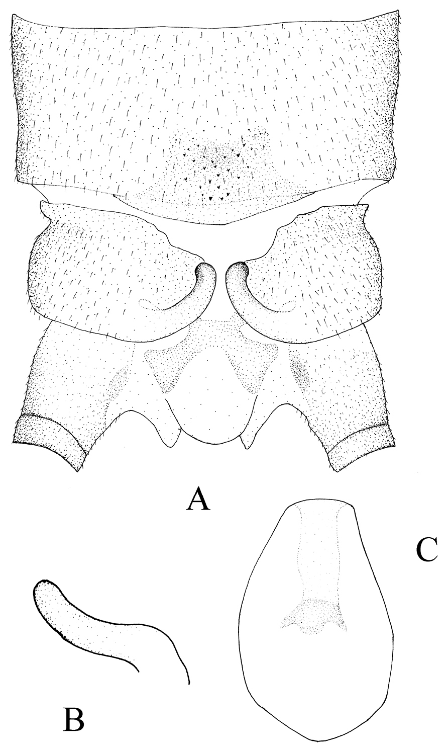
Kamimuria guangxia Li & Wang, sp. n. (male). A Terminalia, dorsal view B Hemitergal process, lateral view C Aedeagus before eversion, ventral view.

Kamimuria guangxia Li & Wang, sp. n. (male). A Aedeagus, dorsal view B Aedeagus, lateral view.
Unknown.
The specific epithet refers to Guangxi autonomous region where the type specimen was collected.
China (Guangxi).
The male of Kamimuria guangxia is characterized by the hemiterga being slightly swollen apically. The aedeagal sac is membranous, medially constricted, apex expanded, heart-shaped and mostly covered with fine to tiny spines (Figs 1C, 2B). The new species is similar to Kamimuria atra Sivec & Stark, 2008, a species known both from Vietnam and Thailand, in general body color and features of terminalia, but tergum 8 of the new species has no sensilla basiconica patch.
urn:lsid:zoobank.org:act:151A4084-8B37-424B-BD9A-8F8CB4A5857A
http://species-id.net/wiki/Neoperla_mesostyla
Figs 4–5Holotype: male, China: Guangxi autonomous region, Mt. Jiuwanshan, Jiuren Station next to Rongjiang River, 950–1150 m, 25.0673N, 109.2563E, light trap, 3 Aug. 2003, Zhang Li-Li. Paratype: 1 male, same data as holotype.
Forewing length ca. 14.8 mm. General body color brown. Distance between ocelli nearly as wide as diameter of ocellus. Head slightly wider than pronotum, with a small triangular interocellar patch and another black triangular patch on frons (Fig. 4A); compound eyes dark; antennae dark brown. Pronotum pale brown with darker anterior and median stripes (Fig. 4A); wing membrane pale brown, veins dark; legs yellowish brown, distal fourth of femora, basal fifth of tibiae and tarsi darker (Fig. 4C).

Neoperla mesostyla Li & Wang, sp. n. (male). A Head and pronotum, dorsal view B Terminalia, dorsal view C Hindleg (part of tarsi in this leg missing), lateral view D Aedeagus before eversion, lateral view E Aedeagus, lateral view.
Process of tergum 7 forming a large subquadrate plateau, mostly covered with dense sensilla basiconica patches but with few sensilla basiconica at margins (Fig. 4B). Tergum 8 with an upcurved tongue-shaped process, with sparse ventral tiny spines. Tergum 9 without sensilla basiconica patches. Hemitergal lobes slender and curved laterally near midlength (Fig. 4B). Aedeagal tube plump (length 3× width at basal bulb), ventrally with a mesal bifurcate lobe bearing a basal common stem, dorsal surface heavily sclerotized, the pigmentation slightly expanded mesolaterally, membranous sac 1.5× as long as tube and gradually curved ventrad to form a loop; sharp to stout small spines occur along dorsal surface toward apex (Figs 4D, E, 5).

Neoperla mesostyla Li & Wang, sp. n. Male aedeagus, lateral view.
Zwick & Sivec defined the bulb side of the tube as being ventral, however, the sac of the new species forms a loop so that the spines of apical half of the dorsal surface are actually ventrally directed; we herein discuss dorsal or ventral surfaces of the sac as if it were straight, as such in Neoperla flagellata Li & Murányi (
Unknown.
The specific epithet refers to the mesal position of the bifurcate lobe on the ventral surface of the aedeagal tube.
China (Guangxi).
The new species appears to belong to a well differentiated subgroup of the montivaga species group (
The new species is characterized by the aedeagal tube being short (≤3× width of bulb) and the ventral, bifurcate process being midlength on the tube. Additionally, the apical half of the sac is clothed with heavy spinules on the dorsal surface. Neoperla diehli has an elongate slender aedeagal tube (length ≥4× tube width), the ventral processes originate at the apex of the tube, and the heavy spinules are restricted to the tip of the sac (see Fig. 21 in
We acknowledge the partial support of our research by the National Natural Science Foundation of China (No. 31000977).