


(C) 2012 Eduardo Suárez-Morales. This is an open access article distributed under the terms of the Creative Commons Attribution License 3.0 (CC-BY), which permits unrestricted use, distribution, and reproduction in any medium, provided the original author and source are credited.
For reference, use of the paginated PDF or printed version of this article is recommended.
During a survey of the zooplankton community of Bahía Amuay, Venezuelan Caribbean, specimens of an undescribed species of Caligus Müller were collected. It resembles Caligus xystercus Cressey and Caligus ocyurus Cressey, both known only from the Caribbean Sea. The new species can be distinguished from these and other congeners by a combination of characters including the armature of legs 1 and 4, but mainly by its unique female genital complex. This is the first species of Caligus described from Venezuela. The species is described in full and a key to the species of the genus recorded in Venezuela is provided.
Bahía Amuay, marine zooplankton, crustaceans, biodiversity
The siphonostomatoid copepods of the genus Caligus Müller, 1785 are one of the most diverse and representative crustacean parasites of teleost fish (
A recent (February 21–24, 2012) biological survey of the planktonic fauna was carried out at the Bay of Amuay, a coastal system on the northern coast of Venezuelan Caribbean coast, which is also an important oil extraction site with pollution problems (
Adult female and male individuals of a caligid copepod of the genus Caligus were recovered from plankton samples obtained from Bahía Norte, a shallow coastal system which is part of the Bay of Amuay (11°46'32"N, 70°13'51"W), in the northwestern coast of Venezuela. The plankton fauna from this area was surveyed during several days (February 21–24, 2011) by performing horizontal hauls with a standard plankton net (0.5 mm mesh size, 0.3 m mouth diameter). Specimens were fixed shortly after collection in 70% ethanol. Specimens of Caligus were sorted from these samples and processed for identification by transferring them to glycerol and then pure glycerine. Drawings were prepared using a camera lucida mounted on an E-200 Nikon compound microscope. Terminology of the body parts and appendages follows
urn:lsid:zoobank.org:act:40EC2919-0E77-4150-B670-C74AC8C3CD0A
http://species-id.net/wiki/Caligus_evelynae
Figs 1–3Holotype female, collected February 21, 2011 by D. Arocha, D. Querales and F. Cancines, Bay of Amuay, Venezuela. Specimen undissected, ethanol-preserved, vial (ECO-CHZ-07565). Allotype male, same date, collector, and site, undissected, ethanol-preserved, vial (ECO-CHZ-07566).
Bay of Amuay, Venezuelan Caribbean, 11°46'32"N, 70°13'51"W, plankton.
Unknown.
Body shape as shown in Fig. 1A, with cephalic shield ellipsoidal with curved lateral margins. Total length 2.55 mm, greatest width 1.1 mm (measured at widest part of cephalothorax). Cephalothorax comprises more than half total length (1.33 mm). Genital complex longer than wide (1.2 x 0.71 mm) with irregularly undulated outer margins and rugged ventral and dorsal surfaces; posterolateral region protruding posteriorly. Abdomen subquadrate, about as long as wide, genital complex approximately 3.4 times longer than abdomen. Caudal rami subrectangular about 1.2 times longer than wide, armed with 3 long terminal, one small outer and one small inner pinnate setae (Fig. 2I). Lunules spaced by the length of about 1.5 times the lunule diameter.
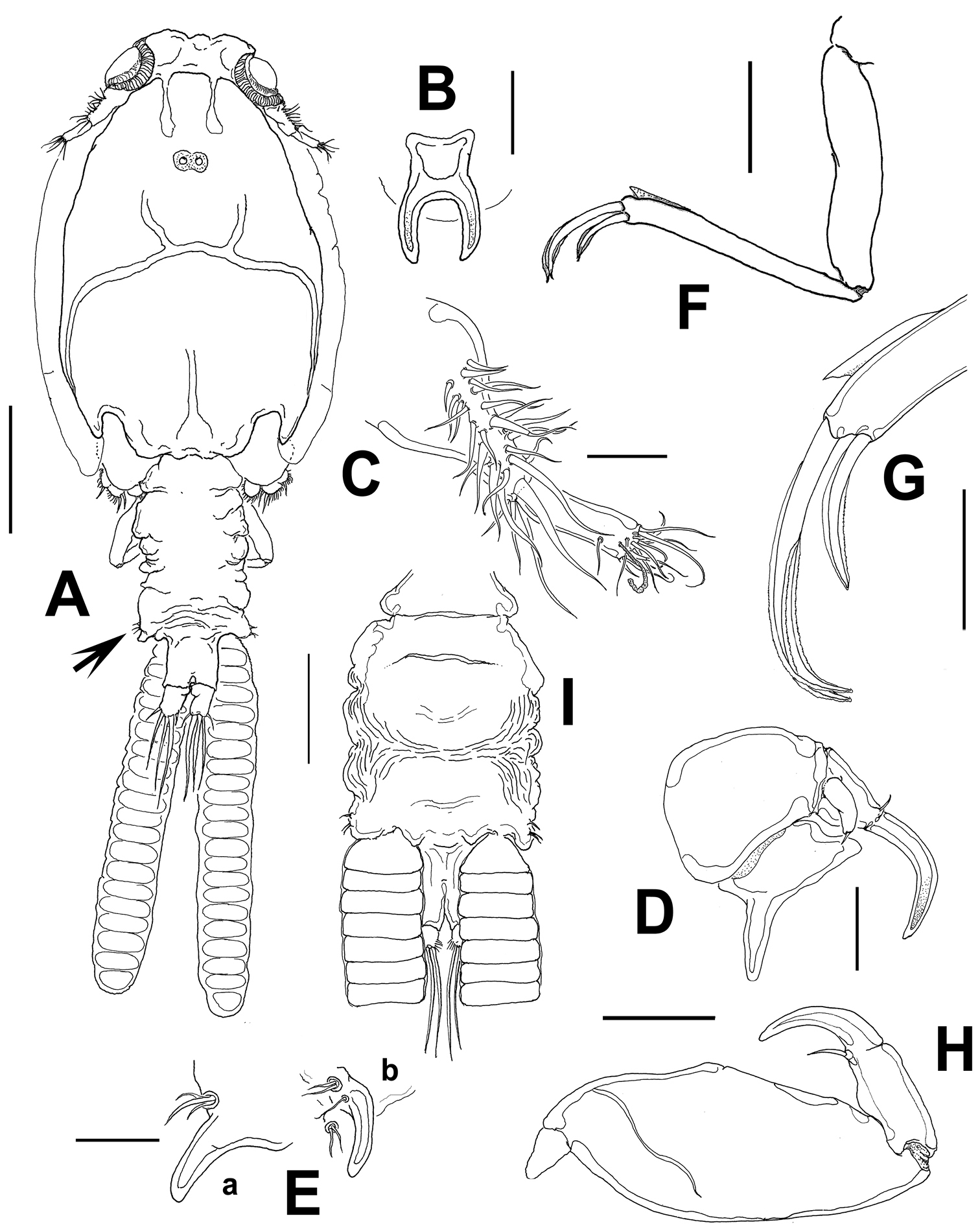
Caligus evelynae sp.n., adult female from Venezuela: A habitus, dorsal view B sternal furca, ventral view C antennule D antenna E postantennal process (b) and maxillule (a) F maxilla G detail of calamus and canna H maxilliped I genital complex and abdomen, ventral view. Scale bars: A, I=0.5 mm, B–F, H =0.1 mm, G=0.05 mm.
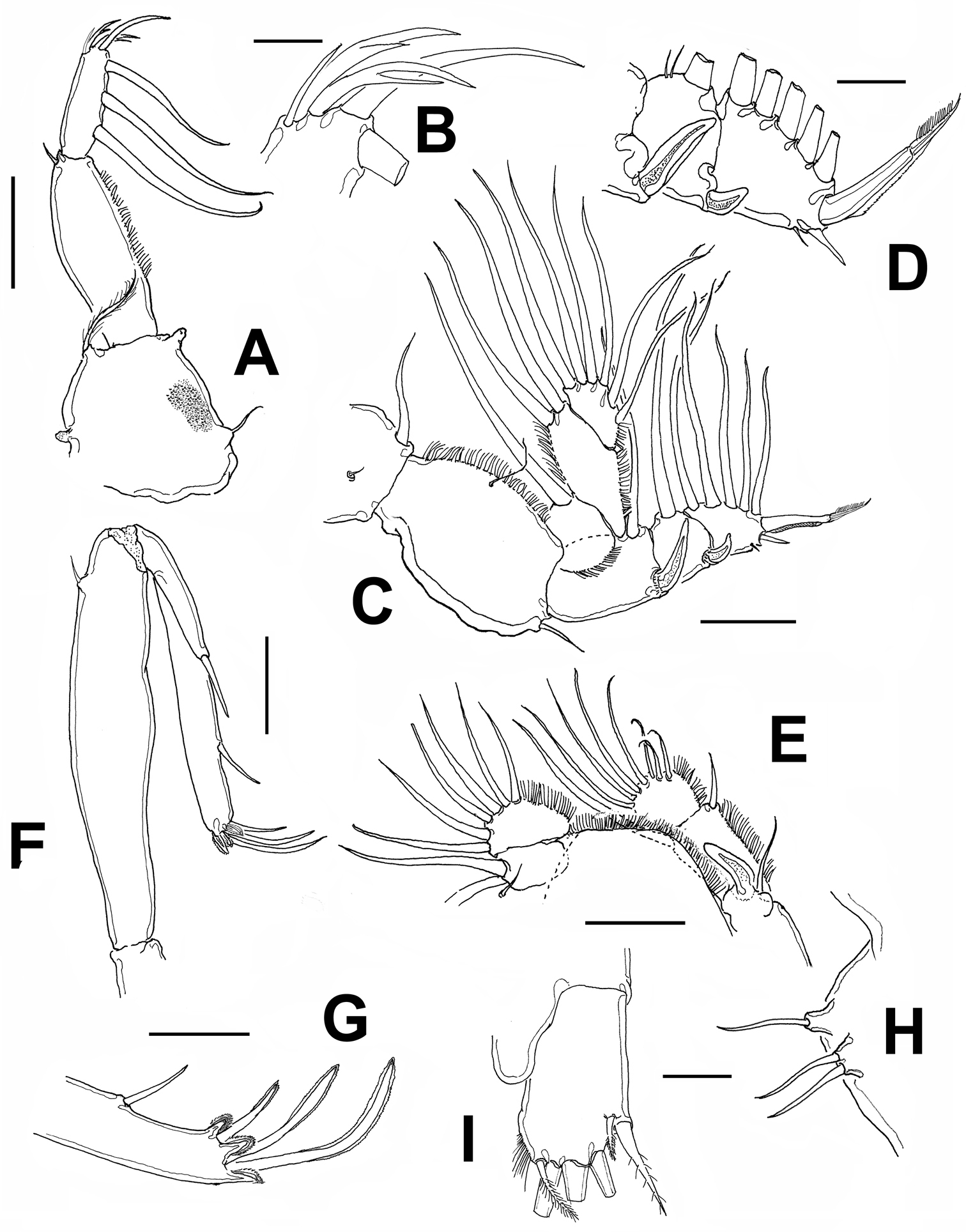
Caligus evelynae sp.n., adult female from Venezuela: A first leg B detail of distal elements of first leg C second leg D detail of exopodal segments of second leg E third leg F fourth leg G detail of terminal elements of fourth leg H fifth leg I caudal ramus, dorsal. Scale bars: A, C, F, E=0.1 mm, B, D, G, H, I=0.025 mm.
Antennule (Fig. 1C) with the usual structure found in Caligus, 2-segmented, proximal segment distinctly longer than distal segment, armed with 22 plumose setae. Distal segment bearing 12 setae (1 of which is subdistal) plus two aesthetascs.
Antenna (Fig. 1D) claw recurved at right angle near tip with small, proximal accessory process; posterior process heavily sclerotized, pointed, but not sharply so. Postantennal process (Fig. 1Eb) sickle-shaped, with rounded tip, with two basal papillae, proximalmost armed with three setae, the other with a single, branched seta. Another papilla with two setae located nearby on sternum.
Maxillule represented by bluntly pointed subtriangular process and basal papilla bearing three small setae (Fig. 1Ea). Maxilla (Figs 1F, G) 2-segmented, brachiform; proximal segment (lacertus) unarmed; distal segment (brachium) slender, with subdistal flabellum on outer margin. Terminal elements, calamus and canna unequally long, the latter about half the length of the former. Maxilliped (Fig. 1H) robust, without protrusions in basal region; subchela about one-half length of basal segment. Terminal claw slightly shorter than shaft, armed with short proximal seta.
Sternal furca (Fig. 1B) tines slightly incurved, membranous on outer margin and longer than base. Leg 1 (Fig. 2A) coxa with patch of very fine spinules in addition to long setulated outer seta and short inner seta. Mammiliform papilla on distal position of segment. First exopodal segment with row of short hair-like elements. Last exopod segment bearing 3 medial pinnate setae and 2 terminal spines, each with accessory process, with additional medial distal seta; usual small seta on outer terminal corner not seen (Fig. 2B).
Leg 2 (Fig. 2C) coxa small, with long plumose seta on inner margin and setule-bearing papilla on middle-outer surface. Basipodite robust, with small seta on outer edge and a setule-bearing papilla on middle inner margin. First exopodal segment bearing spine reaching across second segment to proximal margin of third segment; second segment with much shorter spine recurved on outer margin, spine reaching to middle of last segment. Distal segment with short spine on outer margin plus one short setule. Terminal spine sclerotized, articulated, as long as distal segment (Fig. 2D). All setae on medial margins of all segments pinnate.
Leg 3 (Fig. 2E) exopod first segment with stout, slightly recurved, terminal spine with thin flange on outer lateral margin nearly reaching to third segment; setae as in figure and typical of genus; second endopodal segment with single inner pinnate seta, second segment with 6 pinnate setae.
Leg 4 (Figs 2 F, G) uniramous, brachiform; exopod 2-segmented. Protopod with short plumose seta in distal outer margin. First exopodal segment with terminal seta not reaching base of middle lateral seta of last segment. Second exopodal segment bearing 3 unequally long setae, outermost shortest and medial longest; each with pecten at base.
Armature of rami of legs 1–4 as follows (Roman numerals indicating spines and Arabic numerals, setae):
| exopod | endopod | |
| Leg 1 | 1-0; III, 4 | vestigial |
| Leg 2 | I-1; I-1; II, 5 | 0-1; 0-2; 6 |
| Leg 3 | I-0; I-1; III, 4 | 0-1; 6 |
| Leg 4 | I-0; I, III | absent |
Leg 5 represented by two small papillae on posterolateral corner of genital complex, one armed with two setae, the other with one seta (arrowed in Fig. 1A, Fig. 2H).
Body (Fig. 3A) larger than female, 3.1 mm long excluding setae on caudal rami. Cephalothoracic shield roughly ovoid in shape, 1.78 mm long and 1.35 mm wide (excluding marginal hyaline membranes: 0.07 mm). Frontal plates well developed and carrying moderately large lunules separated by 1.5 the diameter of a lunule; free margin of thoracic zone projecting slightly beyond tips of lateral zones; sinuses moderately deep. Fourth pediger separated from genital complex, roughly hexagonal in shape, about twice longer than wide. Genital somite subrectangular. Abdomen 1mm long, represented by two somites; proximal somite subquadrate, anal somite distinctly longer than wide. Caudal ramus subrectangular, slightly longer than wide, bearing 3 short (one inner, two outer) setae, and 3 long terminal setae. Inner margin naked.
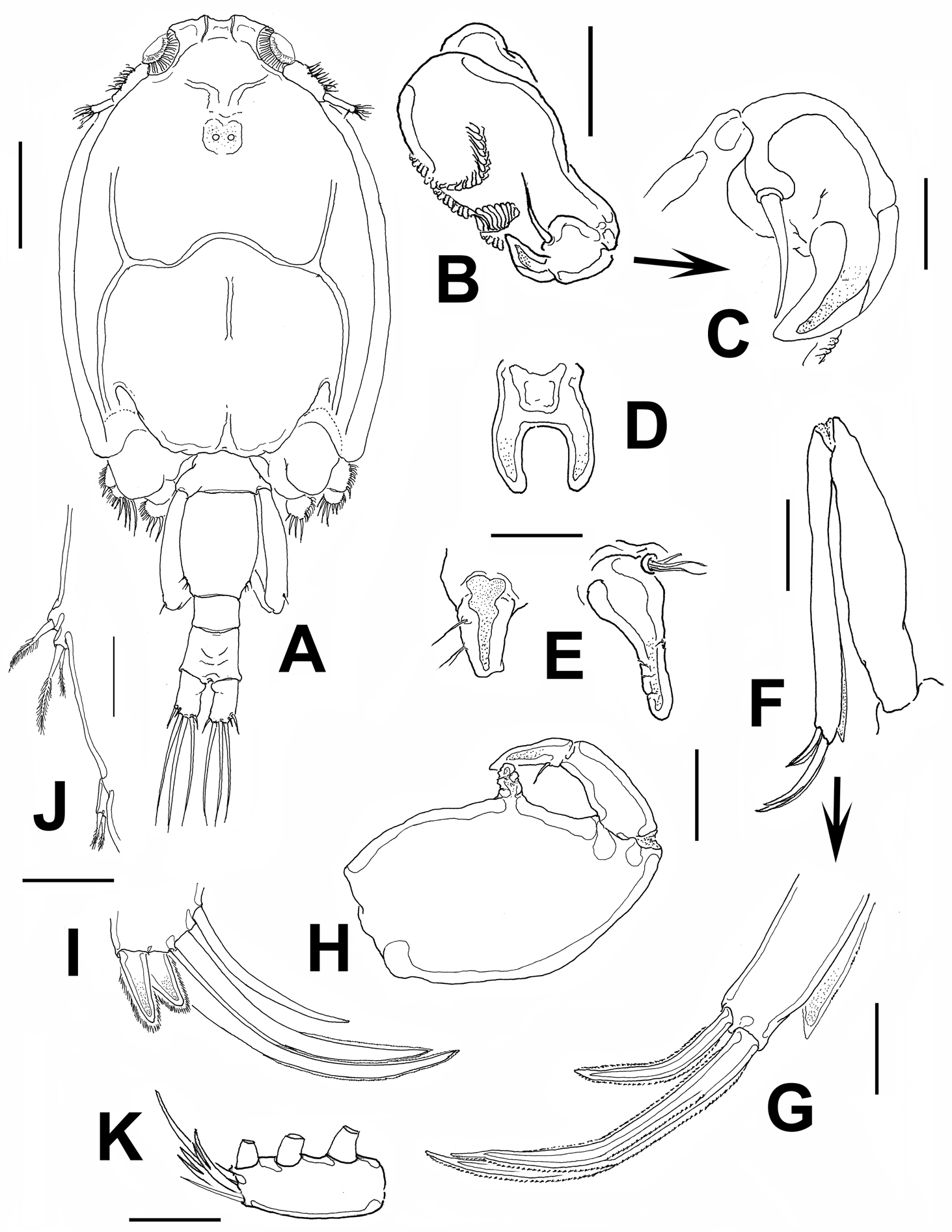
Caligus evelynae sp.n., adult male from Venezuela: A habitus, dorsal view B antenna C detail of distal part of antenna D sternal furca, ventral view E postantennal process and maxillule F maxilla G detail of calamus and canna H maxilliped I fourth leg, detail of distal elements J fifth and sixth legs K first leg, distal segment of exopod. Scale bars: A=0.5 mm, B, D–F, H=0.1 mm, C, G, I, J=0.03 mm; K=0.07 mm.
Antennule as in female.
Antenna (Fig. 3B, C) 3-segmented; proximal segment slender, unarmed, second segment largest, armed with 3 corrugated pads, 1 along inner margin, 2 transverse on outer surface; terminal segment smallest, armed with single basal seta and short, robust hook.
Sternal furca as in female, tines slightly more robust (Fig. 3D). Postantennal process short, distally truncate, armed with one proximal and two medial setae (Fig. 3E).
Maxillule (Fig. 3E) longer than in female, comprising distally blunt dentiform process bearing basal papilla armed with one short and two long setal elements.
Maxilla (Fig. 3F, G) 2-segmented; proximal segment (lacertus) unarmed, slightly shorter than brachium; distal segment (brachium) with subterminal hyaline membrane and two terminal unequal elements (canna about half as long as calamus).
Maxilliped (Fig. 3H) 3-segmented, robust, first segment with medial cylindrical protuberance; second and third segments forming a subchela with subterminal seta.
Leg 1 (Fig. 3K), 2, 3, and 4 as in female, except for relatively shorter outermost terminal claw (Fig. 3I).
Leg 5 (Fig. 3J) located on middle of lateral margin of genital complex, represented by two papillae, one (proximalmost) armed with short seta and the other with two short, slender setae. Leg 6 (Fig. 3J) represented by single papilla armed with two short, pinnate seta.
The new species is named after Dr. Evelyn Zoppi de Roa, an esteemed Venezuelan researcher who pioneered much of the zooplankton research in this country and has lead new generations of planktologists during her long career.
This species is closely related to other two species of Caligus described from the Northwestern tropical Atlantic: Caligus ocyurus Cressey, 1991and Caligus xystercus Cressey, 1991, they all share a longer than wide genital complex, a relatively short abdomen, a similar structure and armature of legs 1 with three inner setae on the last segment and four distal elements, two of them with accessory spines. They have also a fourth leg armed with 5 spines; the three terminal spiniform elements are of different lengths, the outermost being longest.
Following
Overall, the main character distinguishing the new species, Caligus evelynae, is the peculiarly strong, irregular undulation of the genital complex; another species with a genital complex bearing undulated margins reported in the region is Caligus undulatus Shen and Li, 1959 (
It is interesting to point out that these three very similar species of Caligus (Caligus xystercus, Caligus ocyurus, Caligus evelynae) appear to be restricted to the Northwestern Atlantic (
| 1 | Leg 4 exopod with three segments | 2 |
| – | Leg 4 exopod with two segments | 4 |
| 2 | Abdomen more than three times as long as cephalothoracic shield | Caligus bennetti Causey, 1953 |
| – | Abdomen about as long as or slightly longer than cephalothoracic shield | 3 |
| 3 | Inner margin of second exopodal segment of leg 1 with three short setal elements, without postero-lateral process on genital complex | Caligus chorinemi Krøyer, 1863 |
| – | Inner margin of second exopodal segment of leg 1 unarmed; rounded, strongly developed postero-lateral processes on genital complex | Caligus productus Dana, 1852 |
| 4 | Abdomen one segmented | 5 |
| – | Abdomen two or three-segmented | 10 |
| 5 | Abdomen short, about 0.2 times as long as genital complex | Caligus atromaculatus Wilson, 1913 |
| – | Abdomen not as short | 6 |
| 6 | Abdomen about 0.6 times as long as genital complex, without postero-lateral processes on genital complex | 7 |
| – | Abdomen as long as genital complex (or even longer), with strongly protruding postero-lateral processes on genital complex | Caligus bonito Wilson, 1905 |
| 7 | Inner margin of second endopodal segment of leg 2 armed with short, slender setae | 8 |
| – | Inner margin of second endopodal segment of leg 2 armed with tooth-like elements | Caligus asperimanus Pearse, 1951 |
| 8 | Genital complex rugose, with undulate lateral margins | Caligus evelynae sp. n. |
| – | Genital complex differently built | 9 |
| 9 | Middle two of the terminal elements of leg 1 with accessory process | Caligus rufimaculatus Wilson, 1905 |
| – | Middle two of the terminal elements of leg 1 without accessory process | Caligus constrictus Heller, 1865 |
| 10 | Abdomen three-segmented | Caligus coryphaenae Steenstrup & Lütken, 1861 |
| – | Abdomen two-segmented | 11 |
| 11 | Claws of leg 4 decreasing in length from the inner to outer margin, inner setae of distal exopodal segment of leg armed with spines | Caligus mutabilis Wilson, 1905 |
| – | Claws of leg 4 with middle and outer elements being equally long and about half the length of inner claw, inner setae of distal exopodal segment of leg setulated | Caligus irritans Heller, 1865 |
We acknowledge the project: “Línea base biológica portuaria del Puerto de Amuay, dentro del programa de aguas de lastre y sedimentos de la República Bolivariana de Venezuela: columna de agua- componente planctónico”, leaded by Dr. Antonio Herrera (Universidad Marítima del Caribe-UMC). Additional support was provided by the Instituto Nacional de los Espacios Acuáticos (INEA). Dulce Arocha (UMC), David Querales and Franklin Cancines (Armada de la República Bolivariana de Venezuela) helped in the process of plankton sampling. The comments and suggestions from two anonymous reviewers greatly contributed to the improvement of a previous version of this work.