


(C) 2010 Nate Hardy. This is an open access article distributed under the terms of the Creative Commons Attribution License, which permits unrestricted use, distribution, and reproduction in any medium, provided the original author and source are credited.
For reference, use of the paginated PDF or printed version of this article is recommended.
Eight new Australian species of Poliaspisare described and illustrated: Poliaspis alluvia sp. n., Poliaspis araucariae sp. n., Poliaspis ceraflora sp. n., Poliaspis naamba sp. n., Poliaspis nalbo sp. n., Poliaspis narunggasp. n., Poliaspis ozothamnae sp. n., and Poliaspis waibenensis sp. n. Two described species are transferred into Poliaspis and are redescribed and illustrated: Lineaspis callitris (Laing) originally described by Laing as a species of Poliaspis, is transferred back into Poliaspis as Poliaspis callitris Laing comb. rev., and Leonardaspis wilga (Leonardi) is transferred to Poliaspis as Poliaspis wilga (Leonardi) comb. n. Descriptions and illustrations are also provided for six of the fourteen previously-named Poliaspis species, including five from Australia: Poliaspis attenuata Brimblecombe, Poliaspis elongata Brimblecombe, Poliaspis exocarpi Maskell, Poliaspis nitens Fuller, and Poliaspis syringae Laing. Both Poliaspis cycadis Comstock and Poliaspis gaultheriae Green become junior synonyms of Poliaspis media Maskell. The species not treated here are Poliaspis intermedia Fuller (the location of the types is unknown and Fuller’s description is inadequate), Poliaspis casuarinicola Lindinger (missing types), Poliaspis incisaTakagi and de Faveri (recently, and well described in Takagi and de Faveri 2011), and the six New Zealand species recently revised by Henderson (2011). In addition, Laingaspis lanigera (Laing), the adult female of which has 8 clusters of perivulvar pores – as in Poliaspis species – is redescribed and illustrated. Lectotypes are designated for Laingaspis lanigera, Poliaspis callitris, Poliaspis exocarpi, Poliaspis media, and Poliaspis wilga. A key is provided to the species of Poliaspis, excluding Poliaspis casuarinicolaand Poliaspis intermedia butincluding Poliaspis incisa and the New Zealand species: Poliaspis chathamica Henderson, Poliaspis floccosa Henderson, Poliaspis lactea (Maskell), Poliaspis media Maskell, Poliaspis raouliae Henderson and Poliaspis salicornicola Henderson.
taxonomy, species descriptions, armored scale insects
Many armored scale insects (Diaspididae) are pests, and armored scales are disproportionately common in invasive faunas. About 2, 500 species of armored scale insects have been described, and ten percent of these (250 spp.) are known to occur in Australia (http://scalenet.info/country_taxon/Australia/Diaspididae/).
Here, these eight new species of Poliaspis as well as six of the fourteen previously-named species are described and illustrated. We transfer two described species into Poliaspis and redescribe and illustrate these. We also redescribe and illustrate Laingaspis lanigera
(Laing), the adult female of which has a similar distribution of
perivulvar pores, but differs substantially in another key
morphological feature. A key to the species of Poliaspis is provided, excluding Poliaspis casuarinicola and Poliaspis intermedia, but including Poliaspis incisa Takagi & De Faveri, recently described from Northern Queensland on mangroves (
Depositories are abbreviated as follows: ASCU, Agricultural Scientific Collections Unit, Orange Agricultural Institute, New South Wales; BMNH, the Natural History Museum, London, UK; NZAC, New Zealand Arthropod Collection, Auckland, NZ; QDPI, Queensland Primary Industries and Fisheries, Brisbane, Queensland, Australia; QMBA, Queensland Museum, Brisbane.
Measurements were made using the measurement tools in
NIS-Elements BR 3.00, SPI (Build 455). For species with ≤ 10 specimens,
measurements were taken from all specimens; for those with >10
specimens, 10 specimens were measured, chosen to represent the range
of host plants and geographic localities present in the sample. The
morphological terms for Diaspididae follow those of
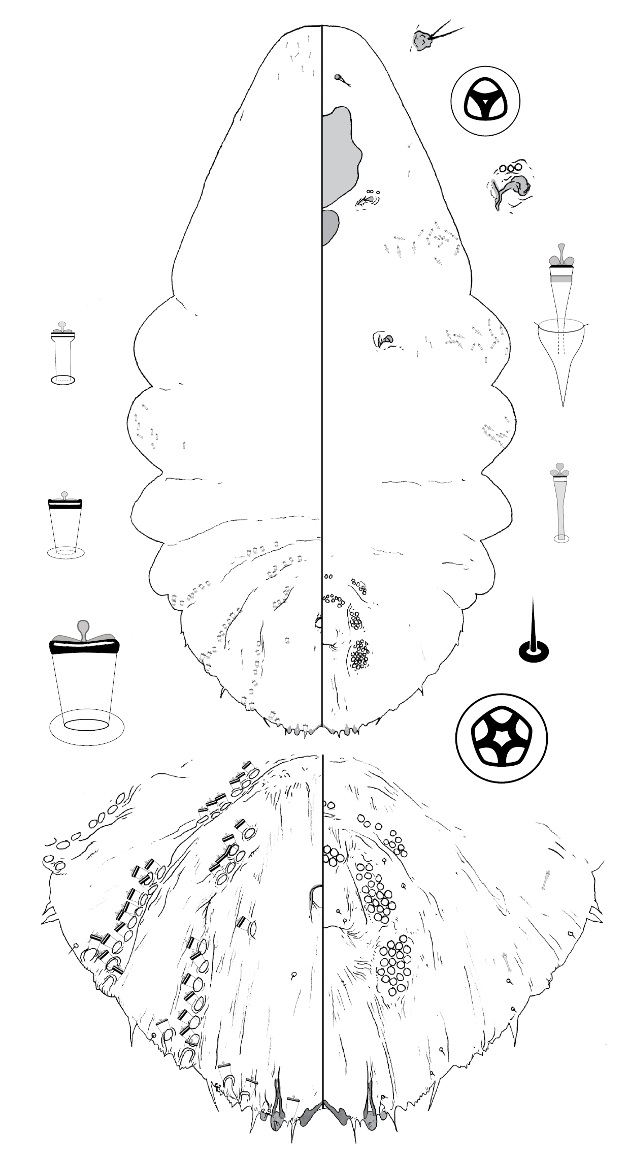
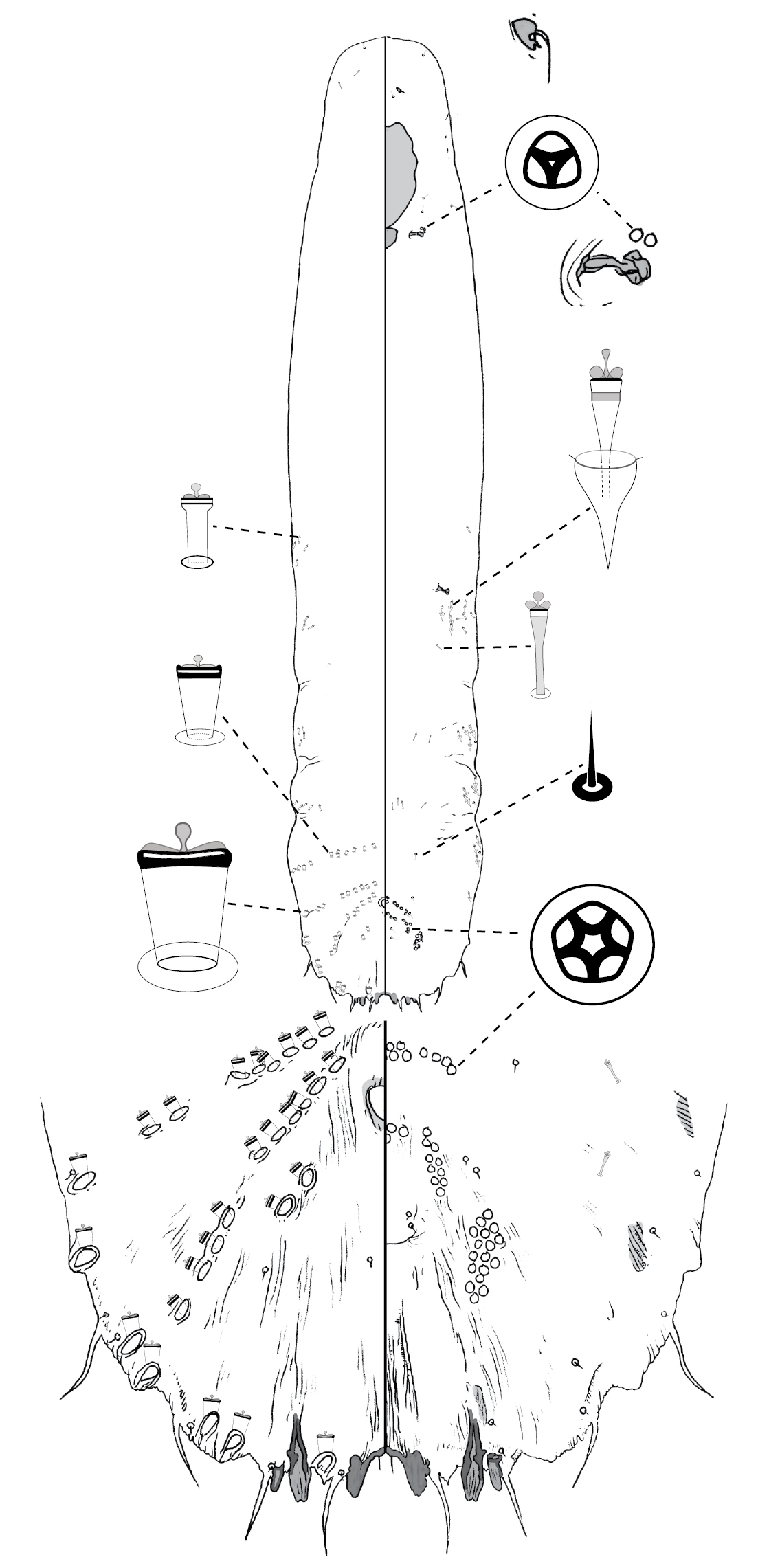
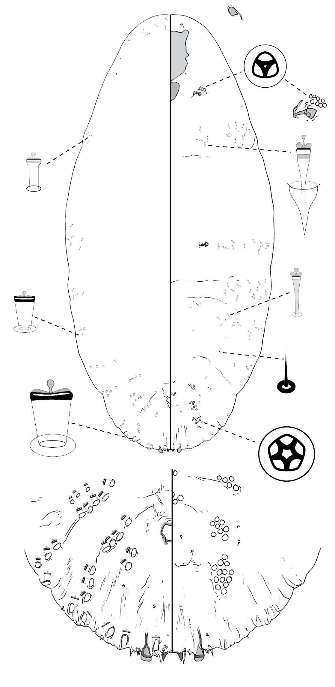
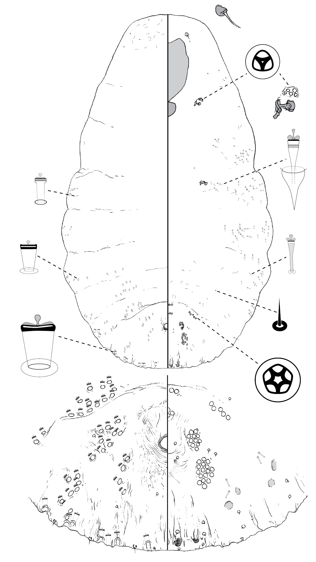
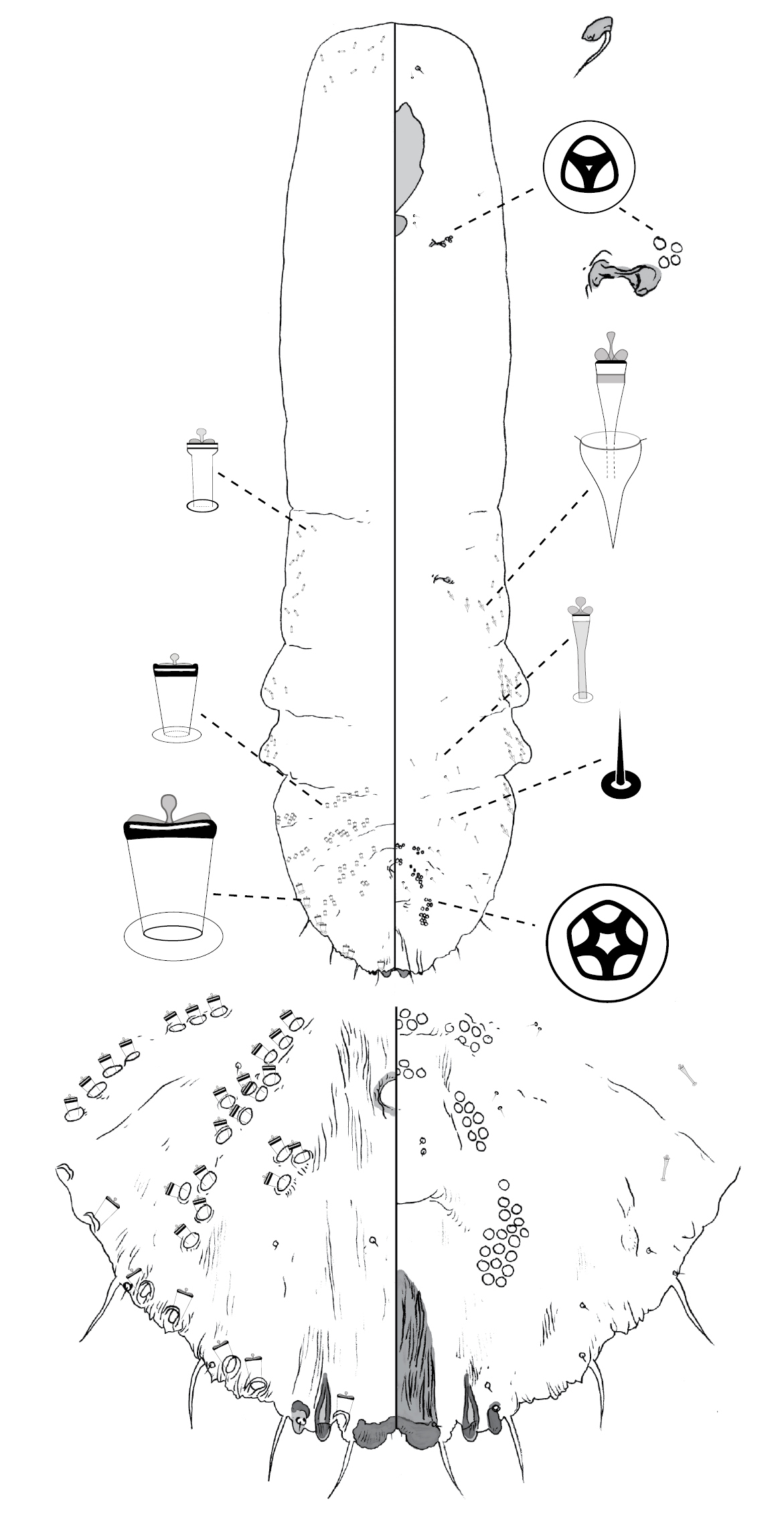
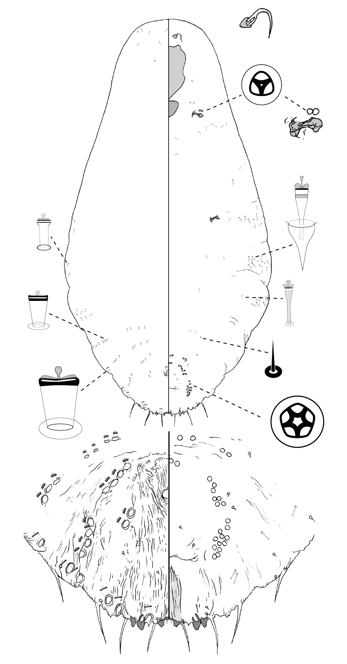
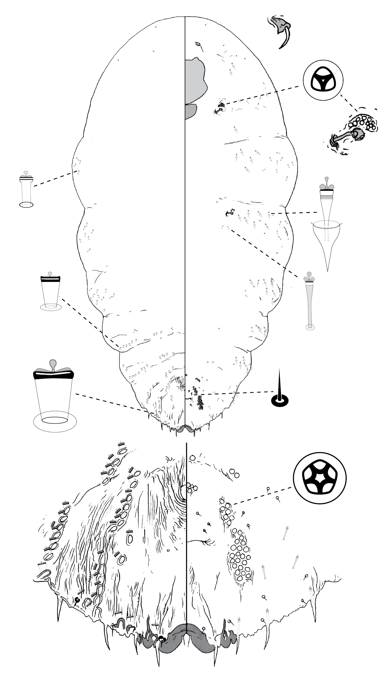
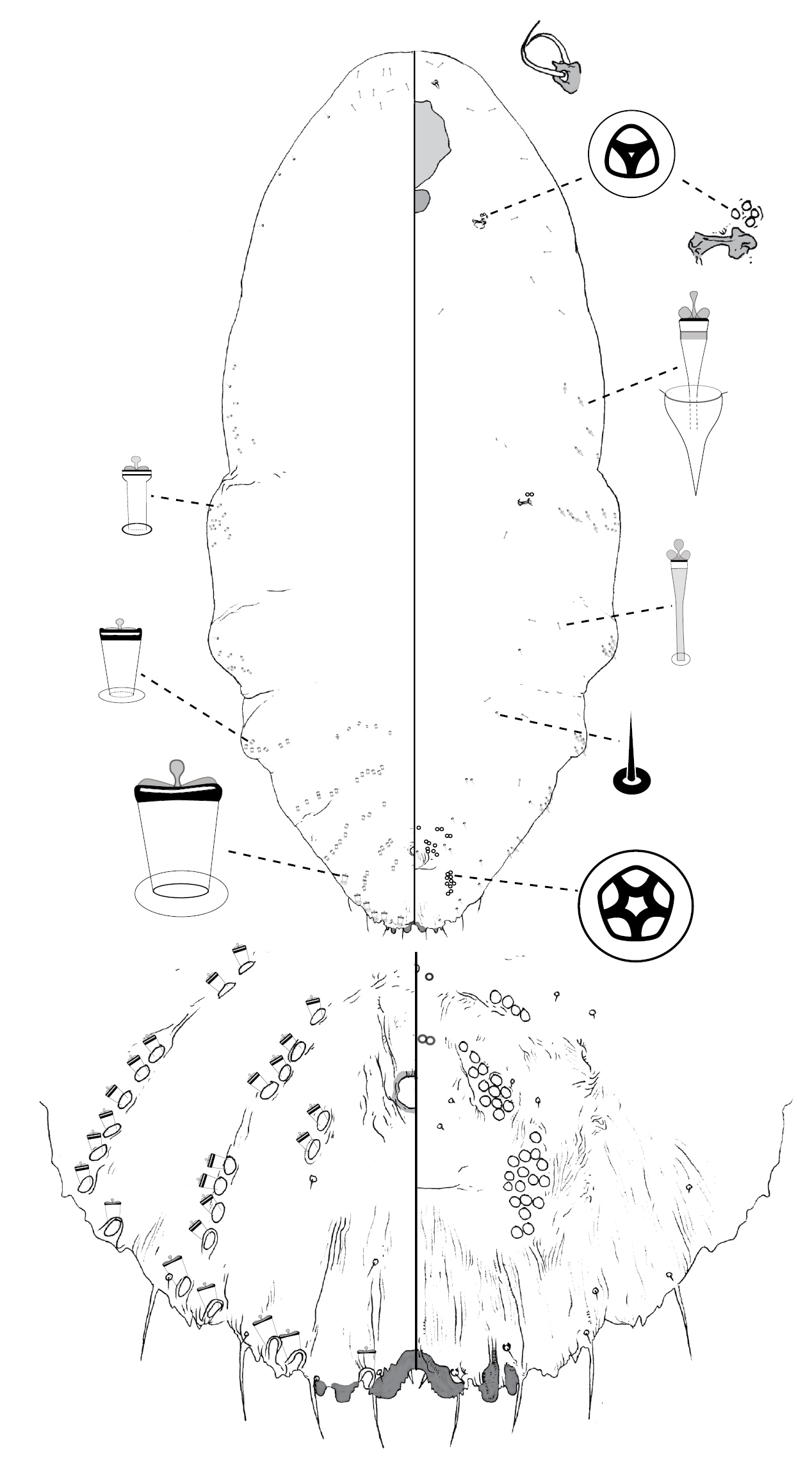
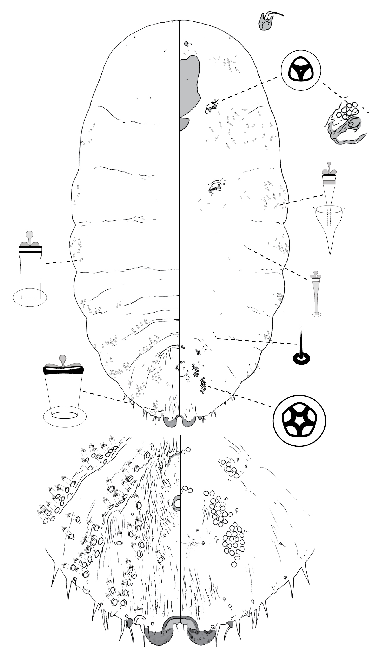
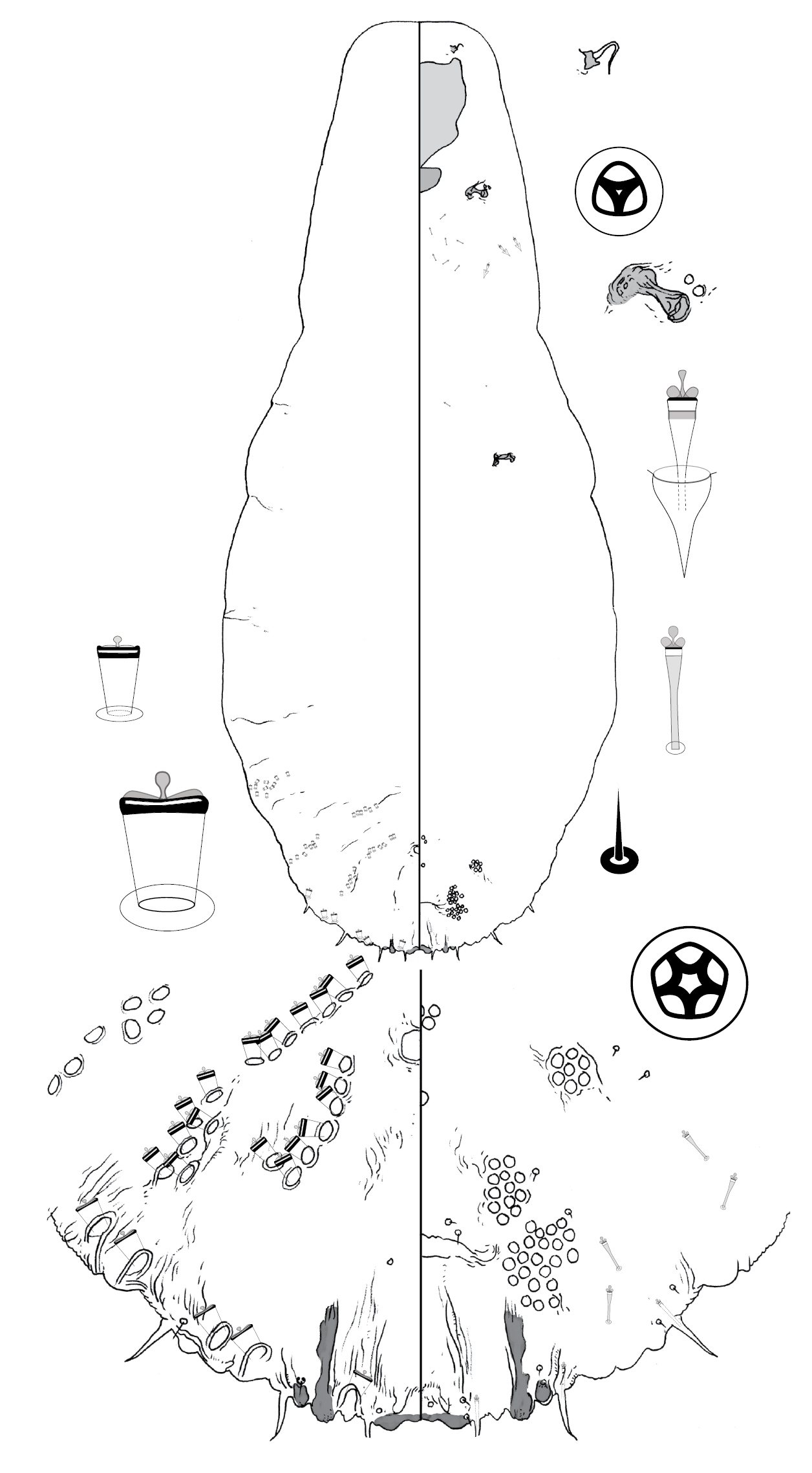
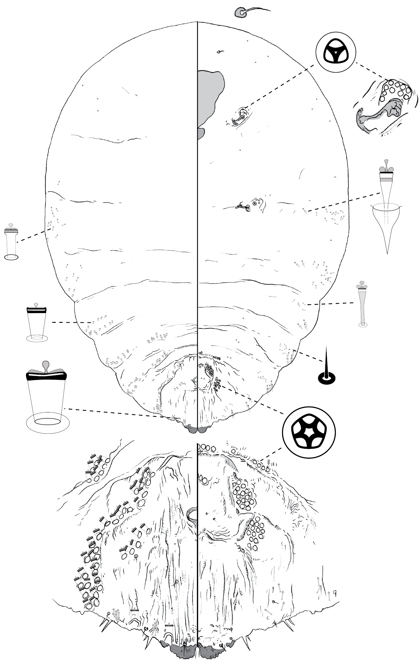
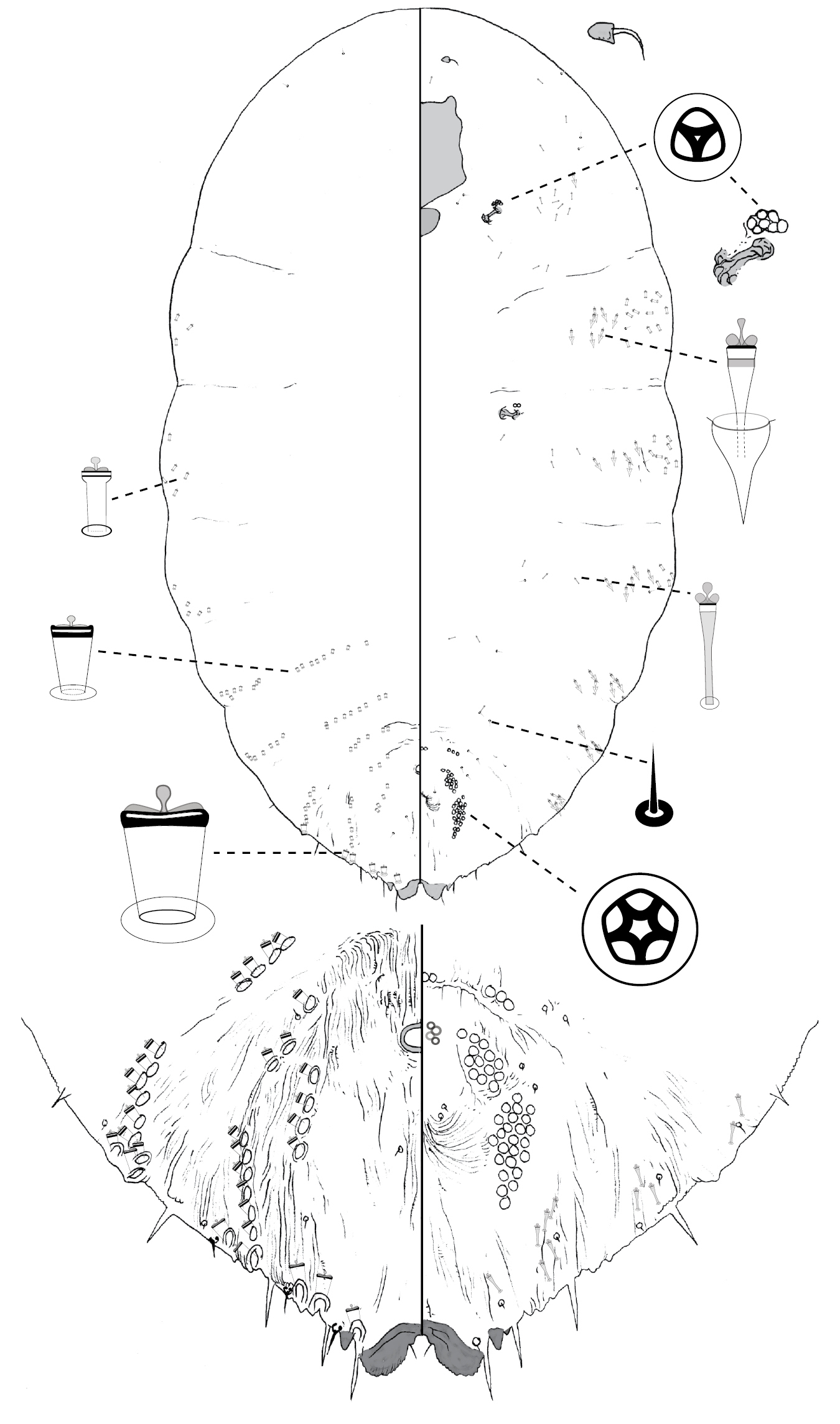
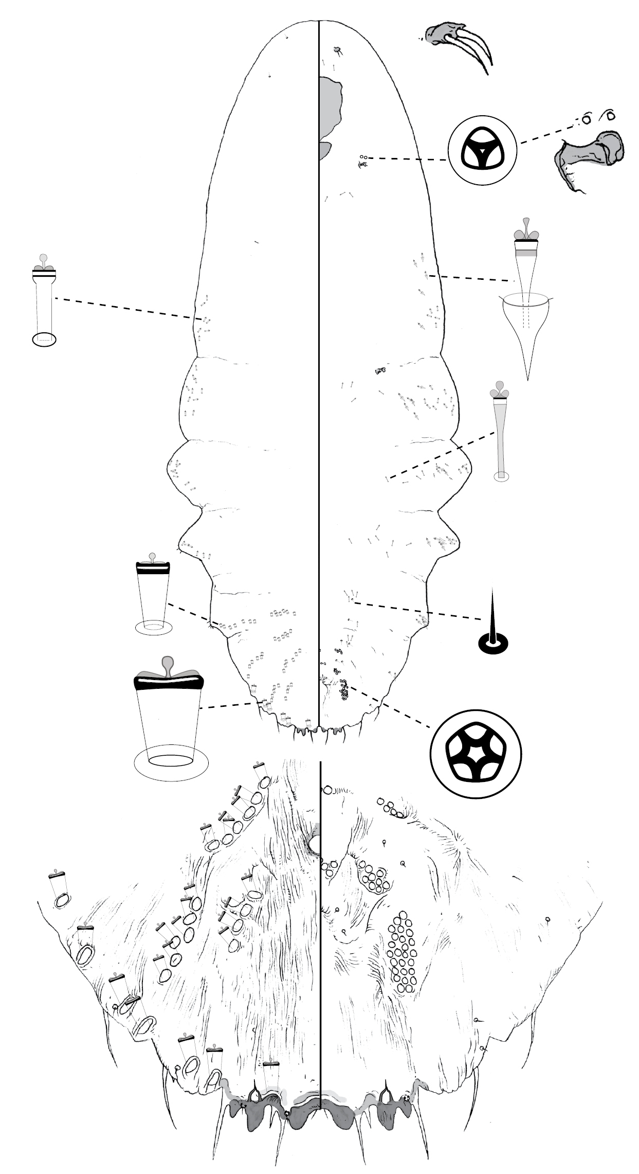
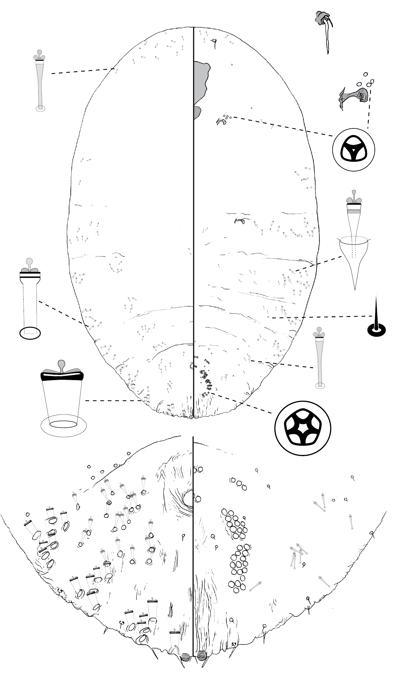
Following the convention for scale insects, each figure displays the dorsal body surface on the left side of the page, and the ventral body surface on the right. Enlargements of diagnostic features are located around the margin of each main figure. Geographic coordinates are provided for each collection location (with a few exceptions). If this information was not part of the original collection data (most cases), approximated coordinates are provided in square brackets. We estimated coordinates via the Google Geocoding API (http://code.google.com/apis/maps/documentation/geocoding/), automating requests via a Python script (available from NBH by request).
Taxonomyhttp://species-id.net/wiki/Poliaspis
Scale cover. Round to elongate-oval, white, flocculent wax sometimes present, exuvia terminal (after
Slide-mounted adult female. Body outline variable: linear, turbinate, pyriform, fusiform or oval, prepygidial abdominal margin weakly incised between segments to strongly lobed. Margin of pygidium rounded; incised between median lobes in some species, not incised in others. Two pairs of lobes in all species except Poliaspis wilga comb. n. (only medial pair) and some New Zealand species (3rd lobe represented by three pointed projections); median lobes zygotic (except in Poliaspis ceraflora), parallel or divergent, apex variable – rounded or pointed; pair of setae between median lobes in most species; second lobes bi-lobed or undivided; basal scleroses present or absent. Simple gland spines present; most species with 1 gland spine on each side of each pygidial segment (other than segment 8), but gland spines may be absent on segment 7 (in area adjacent to lateral margin of medial lobe, e.g. Poliaspis ozothamnae sp. n.), or absent from pygidial segment 5 (Poliaspis ceraflora sp. n., Poliaspis callitris comb. rev.), or 2–6 may be present on each side of segments 5 and 6 (Poliaspis ozothamnae; Poliaspis nalbo sp. n.); length of gland spines variable, from about as long as median lobes to > 5 × length of median lobes. Anus in anterior third of pygidium; opening round. Trilocular pores in cluster near each anterior spiracle, some species also with pores near posterior spiracles. Antenna with 1 or 2 fleshy setae. Perivulvar pores quinquelocular, in 8 groups; 5 groups on abdominal segment 6, and 3 groups on abdominal segment 5. Dorsal ducts 2-barred; ducts on pygidial margin larger than medial ducts in most species; distribution of enlarged marginal ducts: 1 between median and second lobes; 1–2 on segment 6, laterad of second lobes; 2 on segment 5; dorsal ducts (other than those on margin) decreasing in size anteriorly; absent from abdominal segment 7; discrete submarginal and submedial rows of ducts present on any of abdominal segments 2–6: 1–10 submedial ducts present on abdominal segment 6, 4–12 submarginal and 4–15 submedial ducts present on segment 5. Some species with dorsal boss present on submargin of each of abdominal segments 1 and 3. Small ducts similar to dorsal ducts present on ventral submargin. Ventral gland tubercles in marginal / submarginal clusters on thoracic and pre-pygidial abdominal segments. Microducts present on venter, at least along abdominal submargin.
Nearly all other armored scale insect species
have perivulvar pores in no more than 5 clusters, and restricted to
abdominal segment 7. Species of Leucaspis Signoret and Lopholeucaspis
Balachowsky are exceptions to this generalization; more than 5 clusters
of multilocular pores may be present on the abdomen, but the extra
pores occur on the submargin of abdominal segments 6 and 5. More
pertinent exceptions are species in the African genera Rolaspis Hall, Tecaspis Hall, and Dentachionaspis MacGillivray, which have extra perivulvar pores, which occur in the same places as in Poliaspis (
* denotes New Zealand species
| 1 | Gland spines either absent or more than one gland spine on each side of at least some pygidial segments | 2 |
| – | One gland spine on each pygidial segment | 7 |
| 2 | Gland spines absent; marginal macroducts not differentiated from dorsal macroducts; dorsal ducts on pygidium numerous, not arranged into discrete submedial and submarginal clusters; medial lobe pointed, smaller than second lobe | Poliaspis narungga sp. n. |
| – | Gland spines present; marginal macroducts usually larger than dorsal ducts; dorsal ducts on pygidium arranged into distinct, transverse submedial and submarginal clusters; medial lobe either larger or smaller than second lobe | 3 |
| 3 | Median lobes non-zygotic | Poliaspis ceraflora sp. n. |
| – | Median lobes zygotic | 4 |
| 4 | One gland spine on each side of each of abdominal segments 7 and 6; gland spines absent from segment 5; median lobes much smaller than second lobes | Poliaspis callitris comb. rev. |
| – | Two or more gland spines present on each side of each of abdominal segments 6 and 5, or on 5 and 4; median lobes prominent, much larger than second lobes | 5 |
| 5 | Marginal macroducts not much larger than dorsal ducts on pygidium, 1 present on each side of abdominal segment 7, and in a discrete cluster of 2–5 ducts on segment 6 | Poliaspis chathamica Henderson* |
| – | Marginal macroducts clearly larger than dorsal ducts on pygidium, 1 present on each side of abdominal segment 7, and 0 or 1 on segment 6 | 6 |
| 6 | Gland spine present between medial and second lobes; cluster of gland tubercles anterior to anterior spiracles | Poliaspis nalbo sp. n. |
| – | Gland spine absent between medial and second lobes; no cluster of gland tubercles anterior to anterior spiracles | Poliaspis ozothamnae sp. n. |
| 7 | Body shape linear | 8 |
| – | Body shape fusiform, pyriform, or oval, not linear | 9 |
| 8 | Median lobes longer than wide | Poliaspis attenuata Brimblecombe |
| – | Median lobes wider than long | Poliaspis elongata Brimblecombe |
| 9 | Second lobes absent | Poliaspis wilga(Leonardi) comb. n. |
| – | Second lobes present | 10 |
| 10 | Median lobes smaller than second lobes | 11 |
| – | Median lobes larger than or equal to second lobes | 14 |
| 11 | Numerous marginal macroducts crowded along the pygidial margin and submargin, without a clear gap between those on each pygidial segment | Poliaspis floccosa Henderson* |
| – | No more than 2 enlarged macroducts on margin of each pygidial segment, with a clear gap between those on each pygidial segment | 12 |
| 12 | About 7 submedial ducts on each side of abdominal segment 6; median lobes short and broad, pygidial margin between lobes not incised | Poliaspis nitens Fuller |
| – | ≤ Five submedial ducts on each side of abdominal segment 6; median lobes not short and broad, pygidial margin between lobes incised | 13 |
| 13 | Median lobes divergent, each lobe with rounded apex; 1–2 submedial duct on dorsal surface of abdominal segment 6; gland spines ca. 2–3 × as long as median lobes | Poliaspis araucariae sp. n. |
| – | Median lobes parallel, each lobe with pointed or notched apex; 2–5 ducts on dorsal surface of abdominal segment 6; gland spines ca. 4–5 × as long as median lobes | Poliaspis exocarpi Maskell |
| 14 | Median lobes prominent, with more than half total lobe length extending beyond margin | 15 |
| – | Much less than half total lobe length of median lobes extending beyond margin | 17 |
| 15 | One enlarged marginal macroduct on each side of abdominal segment 6; lobules of lobe 2 fused into a single triangular-shaped lobe | Poliaspis salicornicola Henderson* |
| – | Two marginal macroducts on each side of abdominal segment 6; second lobe bi-lobed | 16 |
| 16 | Median lobe zygosis a broad band, posterior spiracles with 2–35 pores, anterior spiracles with 8–150 pores | Poliaspis lactea (Maskell)* |
| – | Median lobe zygosis a narrow strap, posterior spiracles with 1–3 pores, anterior spiracles with 5–19 pores | Poliaspis syringae Laing |
| 17 | Submedial ducts on dorsum of abdominal segment 5 in cluster 2–3 ducts deep | 18 |
| – | Submedial ducts on dorsum of abdominal segment 5 in transverse linear row | 19 |
| 18 | Submedial ducts present on dorsum of abdominal segment 2; ca. 8 submedial ducts on dorsum of abdominal segment 6; median lobes divergent | Poliaspis alluvia sp. n. |
| – | Submedial ducts absent on dorsum of abdominal segment 2; ca. 4 submedial ducts on dorsum of abdominal segment 6; median lobes parallel | Poliaspis incisa Takagi and de Faveri |
| 19 | Second lobes without basal scleroses; marginal macroducts in a group of 2–4 each side of median lobes and a group of 3–6 on each side of segment 6 | Poliaspis raouliae Henderson* |
| – | Second lobes with basal scleroses; 1 marginal macroduct each side of median lobes and 2 marginal macroducts on each side of segment 6 | 20 |
| 20 | Gland spines ca. 1 × length of marginal macroducts | Poliaspis media (Maskell)* |
| – | Gland spines ca. 2–4 × length of marginal macroducts | 21 |
| 21 | Conspicuous duct spur, about as long as lateral lobule of second lobe present between medial and second lobe; usually 4 submedial ducts on dorsal surface of abdominal segment 6; pores usually absent from around posterior spiracle | Poliaspis waibenensis sp. n. |
| – | Conspicuous ducts spur, about as long as lateral lobule of second lobe not present between medial and second lobe; usually 2–3 submedial ducts on dorsum of abdominal segment 6; ca. 2 pores present near posterior spiracle | Poliaspis naamba sp. n. |
urn:lsid:zoobank.org:act:62654309-96F2-413D-BD3C-1DF6CFA5BCBA
http://species-id.net/wiki/Poliaspis_alluvia
Fig. 1Holotype: 1 adult female: Australia, QLD, Mt Whitestone [-27.67, 152.16], ex Loranthaceae, 13.6.1989, M Taylor (ANIC).
Slide-mounted holotype female 952 μm long, body outline pyriform, thoracic and abdominal lobes weakly produced. Pygidium with 2 pairs of lobes; median lobes divergent, with dentate apex; margin between lobes incised to a variable degree; second lobe bi-lobed, each lobule with basal sclerosis, more strongly developed on medial lobule. Gland spines 24–38 μm long, 2–3 × length of median lobes, 1 gland spine on margin of each pygidial segment; pair of setae between median lobes. Dorsal ducts smaller than marginal ducts present in rows; 8 submedial ducts present on segment 6; ca. 9 submarginal and ca. 10 submedial ducts on segment 5; ducts also present on segment 2. Perivulvar pores numerous: ca. 12 posteromedial, ca. 20 posterolateral, ca. 40 posterior, ca. 12 anteromedial, and ca. 5 anterolateral. Trilocular pores in cluster of 7–8 near anterior spiracle; 4 near posterior spiracle. Microducts few to numerous on dorsum of head, scattered anterior to anterior spiracles and mesad of gland tubercles on thorax and abdomen, few or absent on median abdomen. Antenna with 2 fleshy setae.
The relatively large number (ca 8) of submedial ducts on abdominal segment 6, as well as the large number of perivulvar pores (ca 90) can be used to distinguish Poliaspis alluvia from other species of Poliaspis.
The species name is taken from the Latin word alluvio , meaning flood, in commemoration of the flooding of the Brisbane river in January 2011.
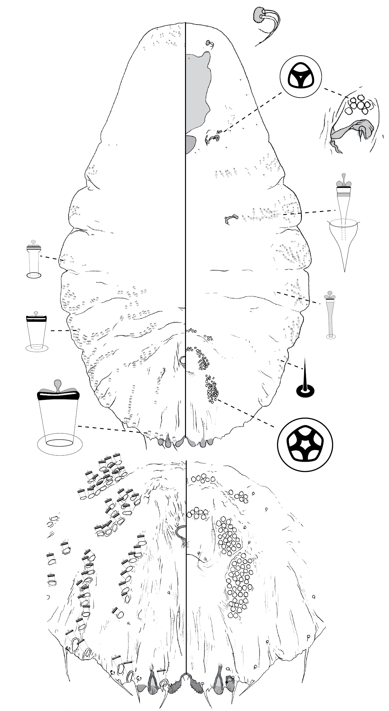
Poliaspis alluvia Hardy & Henderson, sp. n., adult female.
urn:lsid:zoobank.org:act:6A0B48DA-1DC6-4F89-8429-506E76B6EE00
http://species-id.net/wiki/Poliaspis_araucariae
Fig. 2Holotype: female: Australia, QLD, Taromeo [-26.83, 152.12], Araucaria bidwillii, 1.9.1937, A Brimblecombe, 165; 1185/10387 (ANIC).
Paratypes: QLD: 10 adult females: Gallangowan [-27.93, 151.67], ex Araucaria cunninghamii, 15.2.1944, Se/1945 (QDPI); 8 adult females: same data as holotype (QDPI).
Slide-mounted adult female 724–1658 μm long (holotype 1016 μm long), body outline fusiform-pyriform, thoracic and abdominal lobes produced. Pygidium with 2 pairs of lobes; median lobes divergent, connected medially by narrow sclerotic strap, lobes with rounded apex; margin between lobes weakly incised; second lobe bi-lobed, each lobule with basal sclerosis, more strongly developed on medial lobule. Gland spines 12–27 μm long, 2–3 × length of median lobes, 1 gland spine on margin of each pygidial segment; pair of minute setae absent between median lobes. Dorsal ducts similar in size to marginal ducts; present in rows; 1–2 submedial ducts present on segment 6; ca. 6 submarginal and ca. 5 submedial ducts on segment 5. Perivulvar pores: 5–14 posteromedial, 9–20 posterolateral, 23–27 posterior, 1–8 anteromedial, and 7–12 anterolateral. Trilocular pores in cluster of 2–4 near anterior spiracle; absent from posterior spiracle. Microducts scattered on head dorsum, anterior to anterior spiracles and mesad of gland tubercles on thorax and abdomen. Antenna with 2 fleshy setae.
The one or two submedial ducts on abdominal segment 6, in addition to the absence of setae between the median lobes (also absent in Poliaspis callitris, and Poliaspis nitens) can be used to distinguish Poliaspis araucariae from other species of Poliaspis.
The species name refers to the host species: Araucaria cunninghamii and Araucaria bidwillii.
Poliaspis araucariae Hardy & Henderson, sp. n., adult female.
http://species-id.net/wiki/Poliaspis_attenuata
Fig. 3Paratype: QLD. 1 adult female: Yarraman [-26.84, 151.98], ex Croton insularis, 1.9.1948, A Brimblecombe (QDPI).
Slide-mounted paratype female 1644 μm long, body outline linear, abdominal lobes weakly produced. Pygidium with 2 pairs of lobes; median lobes divergent, longer than wide, connected medially by narrow sclerotic strap, each lobe with dentate apex; margin between lobes incised; second lobe bi-lobed, medial lobule with basal sclerosis. Gland spines 18–37 μm long, ca. 2 × length of median lobes, 1 gland spine on margin of each pygidial segment; pair of setae present between median lobes. Dorsal ducts smaller than marginal ducts; present in rows; 2 submedial ducts present on segment 6; 4 submarginal and 5–6 submedial ducts on segment 5. Perivulvar pores: 4 posteromedial, 9–10 posterolateral, 17–18 posterior, 6 anteromedial, and 3–4 anterolateral. Trilocular pores in cluster of ca. 2 near anterior spiracle; absent from posterior spiracle. Microducts scattered on dorsal surface of head, plus medial and submarginal areas of anterior abdominal segments. Antenna with 1 fleshy setae.
Adult females of Poliaspis attenuata are most similar to those of Poliaspis elongata Brimblecombe. Both have elongate, linear bodies. Poliaspis attenuata females can be distinguished on the basis of the longer-than wide, divergent median lobes (wider than long in Poliaspis elongata, with rounded apices).
Poliaspis attenuata Brimblecombe, adult female.
urn:lsid:zoobank.org:act:1C15AF8B-51E2-4C01-B689-1D43E4598E3F
http://species-id.net/wiki/Poliapsis_callitris
Fig. 4Lectotypefemale here designated, 1 of 8 specimens on slide labelled “Australia, VIC, Mallee, on Callitris sp., JE Dixon, no.11, 1919, IBE 1385, EE Green det. ?Chionaspis striata, Poliaspis callitris Laing, sp. n.”The Lectotype is the only un-distorted adult female on slide. (BMNH)
Paralectotypes: (i) the remaining 7 females on the lectotype slide; (ii) 2 adult females on second slide with same collection data and no BM number (BMNH).
Other Material: QLD. 3 adult females: Australia, QLD, Lake Broadwater Conservation park [-27.35, 151.1], on stems of Callitris sp., 19.11.1985, J Donaldson (QDPI); 1 adult female: Southport [-27.97, 153.41], ex Callitris columellaris, 15.8.1953, A Brimblecombe (QDPI); 8 adult females: Southport [-27.97, 153.41], Cupressus macrocarpa, 10.1936, A Brimblecombe (QDPI).
Slide-mounted adult female 581–1188 μm long (holotype 945 μm long), body outline fusiform, without distinct thoracic and abdominal lobes. Pygidium with 2 pairs of lobes; median lobes zygotic, much smaller than medial lobule of second lobe, each lobe with pointed apex; margin between median lobes not incised; second lobe bi-lobed, lateral lobule minute in some specimens (including holotype), medial lobule with strong basal sclerosis. Gland spines 7–11 μm long, about same length as median lobes, 1 gland spine on margin of each of pygidial segments 6 and 7 (i.e. lateral of each lobe), gland spines absent from segment 5; pair of setae absent between median lobes. Dorsal ducts smaller than marginal ducts; present in rows; 2 submedial ducts present on segment 6; ca. 3 submarginal and ca. 4 submedial ducts on segment 5. Perivulvar pores: 2–5 posteromedial, 7–11 posterolateral, 5–13 posterior, 2 anteromedial, and 2–5 anterolateral. Trilocular pores in cluster of 9–15 near anterior spiracle; absent from posterior spiracle. Microducts scattered on dorsal surface of head, mesad of gland tubercles on abdomen. Antenna with 1 fleshy seta.
Adult females of Poliaspis callitris can be distinguished from other species of Poliaspis on the basis of (1) setae between median lobes absent (also absent in Poliaspis araucariae and Poliaspis nitens); and (2) the relatively large number of pores near anterior spiracle (9–15) and no pores near posterior spiracle (other species of Poliaspis having many pores near anterior spiracle have at least a few near posterior spiracle). It shares having only 1 small gland spine on the margin of abdominal segments 6 and 7 with Poliaspis ceraflora , but that species has non-zygotic median lobes.
Poliaspis callitris Laing, comb. rev., adult female.
urn:lsid:zoobank.org:act:1C15AF8B-51E2-4C01-B689-1D43E4598E3F
http://species-id.net/wiki/Poliaspis_ceraflora
Fig. 5Holotype: female: Australia, WA, Perth [-31.95, 115.86], ex Chamelaucium uncinatum, 7.1989, J Donaldson (ANIC).
Paratypes: WA. 5 adult females: same data as holotype (QDPI); 6 adult females: Perth City Council Nursery, ex Melaleuca sp., 8.1973 (ANIC).
Slide-mounted adult female 807–1639 μm long (holotype 1076 μm long), body outline pyriform, thoracic and abdominal lobes weakly produced (undulate). Pygidium with 2 pairs of lobes; median lobes non-zygotic (separate), each lobe with rounded apex; margin between median lobes not incised; second lobe bi-lobed, lateral lobule minute, medial lobule with small basal sclerosis. Gland spines minute, 7–11 μm long, 1 gland spine lateral of each lobe; pair of setae absent between median lobes. Dorsal ducts smaller than marginal ducts; present in non-linear clusters; ca. 4 submedial ducts present on segment 6; ca. 6 submarginal and ca. 8 submedial ducts on segment 5. Marginal ducts: 1 on segment 7, 3 on segment 6, 3–4 on segment 5. Perivulvar pores: 4–5 posteromedial, 9–11 posterolateral, 15–24 posterior, 4–5 anteromedial, and 4–6 anterolateral. Trilocular pores in cluster of 4–5 near anterior spiracle; absent from posterior spiracle. Microducts numerous on dorsal surface of head, scattered mesad of gland tubercles on thorax, in medial and submarginal areas of abdomen. Antenna with 1 fleshy seta.
This is the only species of Poliaspis with non-zygotic median lobes. The two pairs of minute gland spines are also distinctive, although Poliaspis callitris Laing shares the character of possessing only two pairs of small gland spines.
The species name is derived from the Latin words for wax (cera) and flower (floris), in reference to the common name, wax flower, of the host plant genus Chamelaucium.
Poliaspis ceraflora Hardy & Henderson, sp. n., adult female.
http://species-id.net/wiki/Poliaspis_elongata
Fig. 6Paratypes: QLD. 6 adult females: Tugun [-28.14, 153.5], ex Leptospermum whitei, 30.9.1947, A Brimblecombe (QDPI).
Slide-mounted adult female 1323–1886 μm long, body outline linear, with margins of anterior abdominal segments distinctly lobed. Pygidium with 2 pairs of lobes; median lobes zygotic, each lobe wider than long, with rounded apex; margin between lobes not deeply incised; second lobe bi-lobed, lateral lobule small, medial lobule with strong basal sclerosis. Gland spines 22–40 μm long, about 3 × length of median lobes, 1 gland spine on lateral margin of each pygidial segment; pair of minute setae between median lobes. Dorsal ducts smaller than marginal ducts; present in loose rows; 3 submedial ducts present on segment 6; ca. 4 submarginal and ca. 8 submedial ducts on segment 5. Perivulvar pores: 5–8 posteromedial, 8–12 posterolateral, 17–20 posterior, 5–8 anteromedial, and 3–7 anterolateral. Trilocular pores in cluster of 4 near anterior spiracle; absent from posterior spiracle. Microducts numerous on dorsal surface of head, scattered on ventral surface of abdomen. Antenna with 1 fleshy seta.
Adult females of Poliaspis elongata are most similar to Poliaspis attenuata. See comments under Poliaspis attenuata for discussion.
Poliaspis elongata Brimblecombe, adult female.
http://species-id.net/wiki/Poliaspis_exocarpi
Fig. 7Lectotype: female, here designated to preserve nomenclatural stability. On an original slide labelled “Poliaspis exocarpi, adult female, 1891, W.M.M.”. AUSTRALIA, Mordialloc [-38.00, 145.09], near Melbourne, on Exocarpus cupressiformis, by Mr. French (NZAC). Paralectotypes: (i) 1 female, slide label data as above (NZAC); (ii) (not examined) 1 (BMNH); 12 (USNM).
Other material: QLD. 1 adult female: Amamoor [-26.35, 152.68], ex pumpkin, 8.12.1927, H 151a (QDPI); 1 adult female: Amamoor [-26.35, 152.68], ex Cucurbita maxima, 12.12.1927, H 151b (QDPI); 13 adult females: Bamaga [-10.89, 142.39], on leaves of Asteromyrtus lysicephala, 10.9.1983, J Donaldson (QDPI); 3 adult females: Chinchilla [-26.74, 150.63], ex Eremocitrus glauca, 1.12.1981, J Baker (QDPI); 9 adult females: Dalby-Tara-St George Road Junction, on stems of Apophyllum anomalum, 21.11.1985, J Donaldson (QDPI); 4 adult females: Dauan Island [-9.43, 142.53], on leaves, 25.6.1995, J Grimshaw, JFG 2722 (QDPI); 4 adult females: Drillham [-26.64, 149.98], on leaves of Geijera parviflora, 30.4.1953, A Brimblecombe (QDPI); 7 adult females: Emu Vale [-28.23, 152.25], ex Euroschinus falcata, 12.2.1939, A Brimblecombe, 399 (QDPI); 13 adult females: Fletcher [-28.77, 151.87], ex Jacksonia scoparia, 11.1949, (QDPI); 4 adult females: Gabba Island [-9.77, 142.63], on leaves of Exocarpus latiflolius, 13.6.2000, J Grimshaw, JFG 5107 (QDPI); 4 adult females: Gabba Island [-9.77, 142.63], on leaves of Exocarpus latiflolius, 4.6.2003, J Grimshaw (QDPI); 1 adult female: Hopevale [-15.29, 145.11], ex Cycas sp., 6.6.2000, B Waterhouse, JFG 5380 (QDPI); 10 adult females: Imbil [-26.47, 152.7], ex Euroschinus falcata, 10.1936 (QDPI); 7 adult females: Jandowae [-26.78, 151.11], on leaves of Eremocitrus glauca, 26.6.1989, J McAlpine, N5149 (QDPI); 3 adult females: Lake Broadwater Conservation park [-27.35, 151.1], on leaves of Amyema congener, 21.11.1985, J Donaldson (QDPI); 5 adult females: Punsand Bay [-10.87, 142.39], on leaves of Garcinia warvenii, 24.7.2003, J Grimshaw, (QDPI); 7 adult females: Texas [-28.85, 151.17], ex Geijera parviflora, 10.1954, A Brimblecombe (QDPI); 2 adult females: Yarraman [-26.84, 151.98], ex Xanthoxylon brachyacanthum, 6.6.1947, A Brimblecombe, IIE number 1283/10665 (BMNH). VIC. 3 adult females: Dandenong Range [-37.83, 145.35], ex Exocarpus stricta, E Green, BM Reg. Number 1926–415 (BMNH).
Slide-mounted adult female 847–1544 μm long, body outline pyriform to fusiform, with weakly-developed lobes on pre-pygidial abdominal segments. Pygidium with 2 pairs of lobes; median lobes zygotic, parallel, each lobe about as wide as long, with pointed apex, 1–2 notches present in some specimens; margin between median lobes weakly incised; second lobe bi-lobed, lobules similar in size, medial lobule with basal sclerosis. Gland spines 27–43 μm long, about 4–5 × length of median lobes, 1 gland spine on lateral margin of each pygidial segment; pair of setae between median lobes. Dorsal ducts smaller than marginal ducts; present in transverse linear rows; 2–5 submedial ducts present on segment 6; ca. 4 submarginal and ca. 4 submedial ducts on segment 5. Perivulvar pores: 1–3 posteromedial, 6–15 posterolateral, 12–24 posterior, 2–3 anteromedial, and 2–7 anterolateral. Trilocular pores in cluster of 1–8 near anterior spiracle; 0–3 around posterior spiracle. Microducts scattered on dorsal surface of head, plus ventral surface of abdomen. Antenna with 1 fleshy seta.
Poliaspis exocarpi is far and away the most polyphagous and wide spread species of Poliaspis in Australia. There is also a considerable amount of morphological variation present among samples (e.g. the number of submedial ducts on dorsum of abdominal segment 6). The relatively small size of the median lobes (smaller than or equal in size to second lobes) and the relatively long size of the gland spines (up to 43 μm, about 5 × length of medial lobes) are also diagnostic.
Poliaspis exocarpi Maskell, adult female.
http://species-id.net/wiki/Poliaspis_media
Fig. 8Poliaspis media Maskell
Lectotype designated by
Poliaspis cycadis Comstock
Lectotype female here designated, the middle female of 3 on an original slide with two labels: (a) “Poliaspis cycadis Comst. [= undeciphered word], ” (b) “816, Poliaspis cycadis, C.P. & Glye, ” both labels outlined in red and the coverslip ringed in black. USA: Washington DC, in conservatory, ex Cycas revoluta (USNM)
Paralectotypes: (i) the remaining 2 females on the lectotype slide; (ii) 1 female, same collection data (part of type material), subsequently remounted (BMNH); (iii) 2 females, each on a separate slide labelled: Poliaspis cycadis Comst., Type, on Dion edulis, Dept. Agr. D.C.; these subsequently remounted, and with scale cover under separate glass cover slip (USNM).
Poliaspis gautheriae Green
Lectotype female here designated, the female third from the label “TYPE” and marked with an arrow on the slide, in a row of 6 females: on Gaultheria depressa, Botanic Gardens, Edinburgh, Scotland; additional data on envelope: Coll. W. Evans, Oct 1919 (BMNH).
Paralectotypes, all with same collection data: (i) the other 5 females and 4 2nd-exuvia on the lectotype slide; (ii) 7 females, 4 2nd-exuvia and 1 1st-exuvium; (iii) 3 females; (iv) 4 females, on Gaultheria ‘cycadis’ (under glass), November 1919, ex coll. E.E. Green (BMNH).
The type material of Poliaspis cycadis is morphologically inseparable from Poliaspis media.
Both species were discovered at about the same time (1880s) and at
first the host differences (cycads versus wide host range) and
geographic disjunction of North America and New Zealand presented a
conundrum. Examination of the type material of Poliaspis gaultheriae, previously synonymized with Poliaspis cycadis by
Poliaspis media Maskell, adult female.
urn:lsid:zoobank.org:act:4B995A20-0F3B-47C9-97EB-97CC3B0B52DC
http://species-id.net/wiki/Poliaspis_naamba
Fig. 9Holotype: female: Australia, QLD, Nambour [-26.63, 152.96], of Melaleuca sp., 4.2.2005, C Freebaim (QDPI);
Paratypes: QLD. 2 adult females: Bray Park, Brisbane [-27.3, 152.98], ex Melaleuca sp., 6.3.2005, C Freebaim (QDPI); 3 adult females: Cooloola National Park [-26.1, 153.04], on leaves and stems of Monotoca scoparia, 7.4.1987, J Donaldson (QDPI); 3 adult females: Indooroopilly [-27.5, 152.97], on leaves of Melaleuca sp., 6.1989, J Grimshaw (QDPI); 2 adult females: Indooroopilly, 30.10.1998, C Neale (QDPI); 10 adult females: Indooroopilly, of Melaleuca sp., 28.2.2005, C Neale (QDPI); 2 adult females: Kenmore [-27.51, 152.94], on leaves of Melaleuca nodosa, 2.1953, G Smith (QDPI); 7 adult females: Mareeba [-16.99, 145.42], ex Melaleuca bracteata, 12.2004, B Pinese (QDPI); 1 adult females: Mareeba, on leaves of Melaleuca sp., 30.1.1982, J Donaldson (QDPI); 2 adult females: Nambour [-26.63, 152.96], of Melaleuca sp., 4.2.2005, C Freebaim (QDPI); 1 adult females: Yarraman [-26.84, 151.98], on leaves of Guoia semiglauca, 20.3.1952, F Muell. (QDPI).
Slide-mounted adult female 942–1475 μm long (holotype 1475 μm), body outline fusiform to pyriform, with weakly-developed lobes on pre-pygidial abdominal segments. Pygidium with 2 pairs of lobes; median lobes zygotic, divergent, lobes connected via strong sclerosis, each lobe wider than long, with rounded, dentate apex; margin between lobes incised; second lobe bi-lobed, medial lobule larger and with stronger basal sclerosis. Gland spines 19–45 μm long, 2–3 × length of median lobes, 1 gland spine on lateral margin of each pygidial segments; pair of setae between median lobes. Dorsal ducts smaller than marginal ducts; present in rows; 2–4 submedial ducts present on segment 6; ca. 4 submarginal and ca. 4 submedial ducts on segment 5. Perivulvar pores: 1–3 posteromedial, 10–15 posterolateral, 15–23 posterior, 2–4 anteromedial, and 2–5 anterolateral. Trilocular pores in cluster of 3–4 near anterior spiracle; 1–2 near posterior spiracle. Microducts numerous on dorsal surface of head, scattered on ventral surface of abdomen and thorax. Antenna with 1–2 fleshy setae.
Poliaspis naamba is very similar to Poliaspis waibenensis sp. n.Poliaspis naamba adult females can be distinguished from those of Poliaspis waibenensis by (1) lacking a strong duct spur between the medial and second lobes (present in Poliaspis waibenensis); (2) having pores associated with the posterior spiracles (lacking in Poliaspis waibenensis); and (3) with pre-pygidial margin of abdomen only weakly lobed (strongly lobed in P. waibenensis). The two species also have different host associations, with Poliaspis naamba almost always collected from Melaleuca species, and Poliaspis waibenensis from mangrove plants.
The species name is taken from the Aborignal word naamba used in reference to red bottlebrush Melaleuca viminalis. This species has been most often found associated with Melaleuca species.
Poliaspis naamba Hardy & Henderson, sp. n., adult female.
urn:lsid:zoobank.org:act:393A0E21-FA95-4361-8242-CACB6473FEC1
http://species-id.net/wiki/Poliaspis_nalbo
Fig. 10Holotype: female: Australia, QLD, Maleny [-26.76, 152.85], in flower heads of Cryptandra scortechinii, 9.1987, D Hockings (ANIC);
Paratypes: QLD. 4 adult females: same data as holotype (QDPI).
Slide-mounted adult female 685–998 μm long (holotype 998 μm long), body outline elongate oval, margin of thorax and pre-pygidial abdominal segments undulate. Pygidium with 2 pairs of lobes; median lobes large, zygotic, parallel, lobes connected via sclerotic strap, each lobe wider than long, rounded, with dentate apex; margin between lobes incised; second lobe not bi-lobed. Gland spines 17–24 μm long, only slightly longer than median lobes, 1 gland spine on lateral margin of pygidial segment 7 (between medial and second lobes); 2 spines on margin of segment 6; 3 spines on margin of segment 5; pair of setae between median lobes. Dorsal ducts undifferentiated from marginal ducts, except for single larger marginal duct on segment 7; present in clusters (i.e. less organized than rows); 1 submedial duct present on segment 6; ca. 7 marginal-submarginal and ca. 6 submedial ducts on segment 5. Perivulvar pores: 3–4 posteromedial, 14–16 posterolateral, 18–23 posterior, 3–6 anteromedial, and 2–5 anterolateral. Trilocular pores in cluster of 6–8 near anterior spiracle; 2–3 near posterior spiracle. Microducts absent from dorsal surface of head, scattered on ventral surface of head, thorax and abdomen. Antenna with 1 fleshy seta. Cluster of gland tubercles on ventral surface of head anterior to anterior spiracle, in addition to the submarginal / marginal clusters present more posteriorly.
In contrast to many other Australian species of Poliaspis, which are very similar to one another, Poliaspis nalbo is very distinctive. It can be easily recognized by (1) the large, rounded, parallel median lobes; (2) the extra gland spines on the margin of abdominal segments 5 and 6; (3) the cluster of gland tubercles present on the ventral surface of the head (also present in Poliaspis narungga); (4) the absence of microducts from the dorsal surface of the head.
The species name is taken from the name of one of the aboriginal groups that originally populated the type locality Maleny, the Nalbo people.
Poliaspis nalbo Hardy & Henderson, sp. n., adult female.
urn:lsid:zoobank.org:act:5685916A-CDD0-448F-A4BD-D38EF9B8E7D7
http://species-id.net/wiki/Poliaspis_narungga
Fig. 11Holotype: female: Australia, SA, Inneston, Yorke Peninsula [-35.28, 136.94], ex Correa ?reflexa, 1.1975, D Symon (ANIC). Holotype is on slide with 7 additional adult females arranged in two rows. Holotype is in top row, second from left.
Paratypes: SA. 7 adult females: on same slide as holotype (ANIC).
Other material:Australia, 4 adult females, intercepted in quarantine in New Zealand, Auckland, MAF, Lynfield, ex Goeznowia vericosa, 7.7.1997, J. McMillan, (NZAC).
Slide-mounted adult female 1136–1858 μm long (holotype 1552 μm long), body outline elongate oval. Pygidium with 2 pairs of lobes; median lobes zygotic, parallel, lobes connected via narrow sclerotic strap, each lobe ca. as wide as long, pointed, smaller than second lobes, margin between lobes not incised; second lobe bi-lobed, each lobule triangular, with blunt or pointed tip. Gland spines absent; pair of setae between median lobes. Dorsal ducts undifferentiated from marginal ducts; numerous on dorsum of pygidium, not organized into discrete submedial and submarginal clusters. Perivulvar pores: 2–7 posteromedial, 15–28 posterolateral, 30–50 posterior, 4–9 anteromedial, and 7–14 anterolateral. Trilocular pores in cluster of 5–8 near anterior spiracle; absent near posterior spiracle. Microducts absent from dorsal surface of head, scattered on ventral surface of head, thorax and abdomen. Antenna with 1 fleshy seta. Cluster of gland tubercles on ventral surface of head anterior to anterior spiracle, in addition to the submarginal / marginal clusters present more posteriorly.
Poliaspis narungga is the only species of Poliaspis in which the adult females lack gland spines, but gland tubercles are numerous in submarginal areas of the abdomen and thorax, including a cluster anterior to the anterior spiracle. Also distinctive are (1) the lack of differentiation between marginal and dorsal ducts, and (2) the dorsal macroducts on the pygidium not being arranged into distinct submedial and submarginal clusters.
The Narungga people were the inhabitants of the Yorke Peninsula prior to the arrival of Europeans.
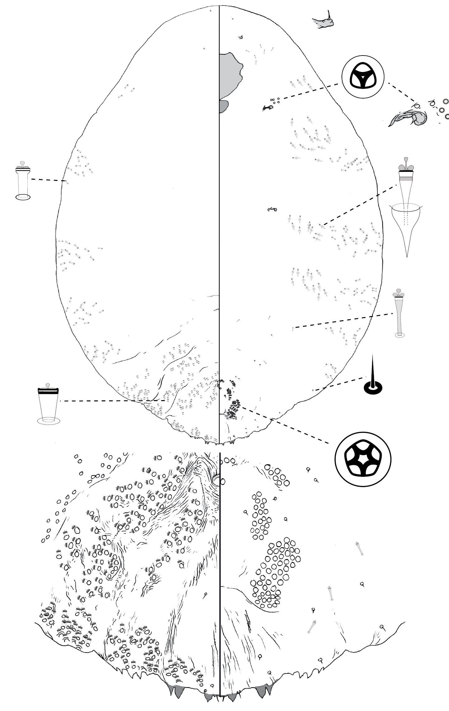
Poliaspis narungga Hardy & Henderson, sp. n., adult female.
http://species-id.net/wiki/Poliaspis_nitens
Fig. 12Dry material: WA. Guilford, ex Daviesia sp., Newman 1912, number 5, WWF 516, ASCT00006373, ASCT00006372; Kalamunda, ex Daviesia sp, Newman 1912, number 2, WWF 517 [corresponds to Froggatt’s collection number 516], ASCT00006371.
VIC. 2 adult females: Sandringham [-37.95, 145.00], ex Exocarpus cupressiformis, C French, IEE 1814; 2 adult females, same coll. data (misidentified as Poliaspis exocarpi) (BMNH). WA. 2 adult females: 4 miles S of Pemberton [-27.47, 153.02], ex stem of Gastrolobium sp., 27.2.1964, SWB (QDPI). WA. 1 adult female mounted from the ASCT0000637 dry material.
VIC. 1 adult female: Dandenong Range [-37.97, 145.24], ex Exocarpus stricta, 2.7.1914, No. 178 G. Brittin Collection (NZAC).
Slide-mounted adult female 1026–1513 μm long, body outline fusiform-pyriform, thoracic and abdominal lobes not produced. Pygidium with 2 pairs of lobes; median lobes short and broad, connected medially by broad sclerosis, each lobe with rounded apex; margin between lobes not incised; second lobe bi-lobed, inner lobule with strong basal sclerosis. Gland spines 17–27 μm long, 2–5 × length of median lobes, 1 gland spine on margin of each pygidial segment; pair of setae between median lobes not observed. Dorsal ducts much smaller than marginal ducts; present in rows; 7 submedial duct present on segment 6; 6 submarginal and 7 submedial ducts on segment 5. Perivulvar pores: 1–4 posteromedial, 7–14 posterolateral, 12–26 posterior, 2–4 anteromedial, and 5–11 anterolateral. Trilocular pores in cluster of 2 near anterior spiracle; absent from posterior spiracle. Microducts scattered on head, posteromedial of anterior spiracle, anteromedial of posterior spiracle, and across abdomen. Antenna with 1 long, curved fleshy setae.
The adult female of Poliaspis nitens can be distinguished from other species of Poliaspis on the basis of the very short and broad median lobes. Three other species treated here have median lobes smaller than the second lobes: Poliaspis callitris, Poliaspis exocarpi and Poliaspis araucariae. In Poliaspis exocarpi and Poliaspis araucariae the body margin between median lobes is slightly incised, and in Poliaspis araucariae the median lobes are strongly divergent. In Poliaspis callitris the body margin is not clearly incised between the median lobes, but each medial lobe is longer than wide and has a pointed apex.
Poliaspis nitens Fuller, adult female.
urn:lsid:zoobank.org:act:1E486663-2B38-4C1A-A2B7-225A5062FA40
http://species-id.net/wiki/Poliaspis_ozothamnae
Fig. 13Holotype: female: Australia, QLD, Brisbane [-27.47, 153.02], ex Ozothamnus diosmifolius, 17.4.1986, N Gough (ANIC).
Paratypes: QLD. 6 adult females: same data as holotype (QDPI). SA. 7 adult females: Second Valley [-35.52, 138.22], ex Pulteneae involucrata, 13.10.1965, HM Brookes (ANIC).
Slide-mounted adult female 809–1256 μm long, body outline turbinate. Pygidium with 2 pairs of lobes; median lobes zygotic, parallel, close-set, lobes connected via narrow sclerosis, each lobe wider than long, apex obtuse-rounded and dentate; margin between lobes not incised; second lobe not bi-lobed, roughly pointed, apex notched in some specimens, close to medial lobe. Gland spines 8–17 μm long, about as long as median lobes, gland spine absent from margin of pygidial segment 7 (between medial and second lobes), 4–7 spines on margin of each of abdominal segments 5–6; pair of setae between median lobes. Marginal ducts: 1 on abdominal segment 7, 1 on segment 6, not differentiated from dorsal ducts on segment 5. Dorsal ducts present in clusters (i.e. several ducts across); ca. 5 submedial ducts present on segment 6; ca. 12 marginal-submarginal and ca. 12 submedial ducts on segment 5. Perivulvar pores: 3–6 posteromedial, 7–15 posterolateral, 10–18 posterior, 4–5 anteromedial, and 3–6 anterolateral. Trilocular pores in cluster of 7–16 near anterior spiracle; 2–8 near posterior spiracle. Microducts absent on dorsal surface of head, scattered on ventral surface of thorax and abdomen. Antenna with 1 fleshy seta. Gland tubercles absent from ventral surface of head anterior to anterior spiracle.
Poliaspis ozothamnae is distinguishable from other species of Poliaspis by having (1) the second lobe close set to medial lobe, without a gland spine near lateral edge of medial lobe; (2) only 2 differentiated marginal ducts; (3) the 2–8 pores near each posterior spiracle; and (4) having multiple gland spines on each pygidial segment other than 8.
The species name is taken from the host name Ozothamnus diosmifolius.
Poliaspis ozothamnae Hardy & Henderson, sp. n., adult female.
http://species-id.net/wiki/Poliaspis_syringae
Fig. 14Lectotype female here designated: Australia, Victoria, Kew, on lilac, C Plumridge, No. 40 (1925), L.B.E. 1413 (BMNH).
Paralectotypes: collection data same as lecototype: 3 females on one slide.
Other material: QLD. 4 adult females: 12 Mile Barramundi Reserve, near Bajool [-23.65, 150.64], ex native citrus, 28.9.2005, R Elder (QDPI); 5 adult females: Darnley Island [-9.58, 143.79], ex Capparis sp., 25.4.1996, J Grimshaw, JFG 3291 (QDPI); 4 adult females: Hannaford [-27.44, 150.04], ex Eremocitrus glauca, 4.7.1979, P Fayden (QDPI);13 adult females: Marmor [-23.68, 150.71], ex Eremocitrus glauca, 10.1955, A Brimblecombe (QDPI); 2 adult females: Yarraman [-26.84, 151.98], ex Capparis nobilis, 8.1947, A Brimblecombe, 2111; 1314/10729 (QDPI); 2 females, UK, Royal Gardens, Kew, England, ex coll. J.W. Douglas, on Cycas revoluta, 1887 (BMNH).
Slide-mounted adult female 453–1662 μm long, body outline oval, margin of thorax and abdomen undulate. Pygidium with 2 pairs of lobes; median lobes large, zygotic, divergent, connected via a narrow strap, each lobe wider than long, apex rounded and dentate; margin between lobes incised; second lobe bi-lobed, lateral lobule minute, each lobule with rounded apex, basal scleroses absent. Gland spines 17–34 μm long, 1–2 × as long as median lobes, 1 gland spine on margin of each pygidial segment; pair of setae between median lobes. Dorsal ducts smaller than marginal ducts, in rows; ca. 4 submedial ducts present on segment 6; ca. 6 submarginal and ca. 3 submedial ducts on segment 5. Perivulvar pores: 2–6 posteromedial, 10–15 posterolateral, 13–24 posterior, 1–6 anteromedial, and 2–4 anterolateral. Trilocular pores in cluster of 5–19 near anterior spiracle; 1–3 near posterior spiracle. Microducts absent on dorsal surface of head, scattered on ventral surface of thorax and abdomen. Antenna with 1 fleshy seta.
Poliaspis syringae is most similar to Poliaspis naamba and Poliaspis waibenensis. Adult females of Poliaspis syringae can be distinguished from those by (1) median lobes much larger than second lobes (similar in size in Poliaspis naamba and Poliaspis waibenensis); and (2) second lobes without basal sclerosis (present in P. naamba and P. waibenensis). The specimens examined here are from Eremocitrus (or native citrus) and Capparis. The specimens from Capparis are larger than those from Eremocitrus (smallest from Capparis 1233 μm, largest from Eremocitrus 1056 μm), have more pores around the anterior spiracle (ca 18 vs ca. 6), and have slightly longer gland spines on the longer side of the range observed among samples from Eremocritus.
Poliaspis syringae Laing, adult female.
urn:lsid:zoobank.org:act:4A9E74E1-428A-4257-9FCE-65A2FDDC1AE1
http://species-id.net/wiki/Poliaspis_waibenensis
Fig. 15Holotype: female: Australia, QLD, Thursday Island [-10.58, 142.22], on leaves of Lumnitzera racemosa, 2.9.2004, B Waterhouse (ANIC).
Paratypes: QLD. 10 adult females: Atherton [-17.27, 145.48], on leaves of Parsonsia straminea, 1.2.1982, J Donaldson (QDPI); 6 adult females: Hammond Island [-10.55, 142.21], on leaves of Rhizophora sp., 29.11.1993, J Grimshaw (QDPI); 7 adult females: Thursday Island, on leaves of Pemphis acidula, 6.9.1983, J Donaldson (QDPI); 5 adult females: Thursday Island, ex mangrove, 16.5.1985, J Donaldson (QDPI); 4 adult females: same data as holotype (QDPI). WA. 3 adult females: Sunday Island [-16.4, 123.19], on leaves of Ficus sp., 13.5.2002, A Williams (QDPI); 3 adult females: Willie Creek via Broome [-17.96, 122.24], ex mangrove, 7.8.2003, A Postle, C Brockway (QDPI).
Slide-mounted adult female 1101–2040 μm long (holotype 2040 μm long), body outline fusiform to pyriform, with weakly-developed lobes on pre-pygidial abdominal segments. Pygidium with 2 pairs of lobes; median lobes zygotic, divergent, lobes connected via broad (more than half width of lobes) sclerosis, each lobe wider than long, with rounded apex; margin between lobes incised; second lobe bi-lobed, ca. as large as medial lobe, medial lobule larger and with stronger basal sclerosis. Gland spines 25–47 long long, 2–3 × length of median lobes, 1 gland spine on lateral margin of each pygidial segment; pair of setae between median lobes. Conspicuous, duct spur present between medial and second lobe, as long as medial lobe. Dorsal ducts smaller than marginal ducts; present in rows; 4 submedial ducts present on segment 6; ca. 6 submarginal and ca. 6 submedial ducts on segment 5. Perivulvar pores: 2–6 posteromedial, 6–11 posterolateral, 15–28 posterior, 0–3 anteromedial, and 4–7 anterolateral. Trilocular pores in cluster of 1–5 near anterior spiracle; absent near posterior spiracle in most specimens (2 present in specimens from Parsonsia). Microducts absent on dorsal surface of head, scattered on ventral surface of abdomen and thorax. Antenna with 2 fleshy setae.
Poliaspis waibenensis is very similar to Poliaspis naamba. See comments under Poliaspis naamba for discussion.
The species name is taken from the Torres Strait Islander name for Thursday Island: Waiben, meaning ‘place of no water.’
Poliaspis waibenensis Hardy & Henderson, sp. n., adult female.
http://species-id.net/wiki/Poliaspis_wilga
Fig. 16Lectotypefemale here designated, female slide labelled: ‘on wilga, Geijera parviflora, Condobolin, 17.x.1900, WW Froggatt (339), BM 1964–4, CIE’ (BMNH).
Paralectotypes: 12 slides with same data as Lectotype (BMNH); 1 slide with 4 females labelled “co-type, Mytilaspis wilga, Green [crossed out and replaced in pencil by ‘Leon.’], ‘Wilga’ Geijera parviflora, NS Wales, Australia, coll. WW Froggett no. 339 [no date].” On cover: “Lepidosaphes wilga Leon., on Geijera, Australia, coll. WW Froggatt, BM 1940, 180, [no collection date].”
Other Material: NSW. 2 adult female: Glenmore via Bourke [-30.09, 145.94], ex Eremophila sp., 10.3.1994, D Sparks (QDPI); 5 adult females: McCatheys, Dunsandle Rds [? ], on leaves of Eremophila deserti, 11.10.1994 (QDPI). VIC. 6 adult females: Merbein [-34.17, 142.05], ex Myoporum sp., 8.3.1948, P26 (QDPI).
Slide-mounted adult female 939–1542 μm long (Holotype 1130 μm long), body outline oval. Pygidium with 1 pair of lobes; median lobes small, zygotic, parallel, lobes connected via narrow sclerosis, each lobe wider than long, rounded; margin between lobes not incised. Gland spines 9–24 μm long, ca. 1–2 × as long as median lobes, 1 gland spine on lateral margin of each pygidial segment; pair of setae between median lobes. Marginal ducts: 1 on abdominal segment 7, ca. 4 on segment 6, ca. 3 on segment 5. Dorsal ducts smaller than marginal ducts; present in clusters (i.e. less organized than rows); 4–6 submedial ducts present on segment 6; 5–7 marginal-submarginal and ca. 9 submedial ducts on segment 5. Perivulvar pores: 3–4 posteromedial, 10–19 posterolateral, 13–27 posterior, 0–4 anteromedial, and 1–8 anterolateral. Trilocular pores in cluster of 2–5 near anterior spiracle; absent near posterior spiracle. Microducts numerous on dorsal surface of head, scattered on ventral surface of thorax and abdomen. Antenna with 2 fleshy setae.
Poliaspis wilga is the only species of Poliaspis to have only the median lobes present. It can also be recognized by the loose groupings of more than 2 marginal ducts on abdominal segments 6 and 7.
Poliaspis wilga (Leonardi) comb. n., adult female.
http://species-id.net/wiki/Laingaspis_lanigera
Fig. 17Lectotypefemale here designated, adult female on slide labelled: Australia, NT, Darwin, on mangrove, GF Hill, BM 1916–225 [with “Type” label] (BMNH).
Paralectotypes: adult female on slide with same data as Lectotype, marked as Paratype [sic] (BMNH); 4 slides of females with same basic data but variously with “Port Darwin on Foreshore, IBE 647, ” “GF Hill no. 682, from type material, BM 1940–180, ” “IBE 151 / GE Hill 682” (BMNH); 6 poor quality single-specimen slide mounts of males, labelled “Australia, Port Darwin, GF Hill, 16.vi.1916, BM 1922–155” – probably part of the type series even though rigistered years later.
Other material: QLD. 5 adult females: Jacky Jacky Creek [-10.94, 142.51], on leaves of Aegiceras corniculatum, 16.1.1998, J Grimshaw, B Waterhouse (QDPI); 5 adult females: New Mapoon [-10.87, 142.39], on stems of Acalypha milkeriana, 14.1.1998, J Grimshaw (QDPI).
Slide-mounted adult female 965–1126 μm long (Holotype 1026 μm long), body outline oval. Margin of pygidium rounded, serrate. Median lobes dinstinct, with pointed apices, rest of pygidial margin densely-packed with gland spines and sclerotic teeth, some of which may be homologous to lobes. Anus in anterior third of pygidium; opening round. Dorsomedial ducts on pygidium longer than marginal and submarginal ducts; present in 2 clusters near anus; ca. 5 ducts in anterior cluster and ca. 3 in posterior one. Trilocular pores in cluster of 2–3 around each anterior spiracle, absent near posterior spiracles. Antenna with 2 fleshy setae. Perivulvar pores quinquelocular, in 7–8 groups; 5 groups on abdominal segment 7, and 2 or 3 groups on abdominal segment 6; 0–4 posteromedial, 5–7 posterolateral, 11–15 posterior, 0–3 anteromedial, and 0–4 anterolateral. Dorsal ducts 1-barred; dense along margin and submargin of pygidial dorsum, plus a few in clusters near anus. Gland tubercles in marginal / submarginal clusters on thoracic and pre-pygidial abdominal segments.
This species has the diagnostic distribution of perivulvar pores found among species of Poliaspis but differs in an important feature: namely, 1-barred ducts arranged in a dense marginal swath on the pygidial dorsum. 1-barred ducts and gland tubercles, which are also present in this species, is an unusual combination among armored scale insect species.

Laingaspis lanigera (Laing), adult female.
This work was supported by an ABRS grant awarded to NBH, and a research investment by the Ministry for Science and Innovation to RCH. We thank Dug Miller (USNM) and Jon Martin (BMNH) for kindly arranging loans of material, and to Jon Martin for also helping with lectotype designations. Penny Gullan located the collection data in WW Froggatt’s notebooks associated with the dry material of P. nitens in ASCU. Thanks also to two anonymous reviewers for helpful criticism on a draft of the manuscript.