ZooKeys 176: 103–121, doi: 10.3897/zookeys.176.2318
Biomineralizations: insights and prospects from crustaceans
Gilles Luquet 1
1 Biogéosciences, UMR 5561 CNRS - Université de Bourgogne, Dijon, France

Corresponding author: Gilles Luquet (gilles.luquet@u-bourgogne.fr)

Academic editor: J. Štrus

received 4 November 2011 | accepted 19 December 2011 | Published 20 March 2011


(C) 2011 Gilles Luquet. This is an open access article distributed under the terms of the Creative Commons Attribution License 3.0 (CC-BY), which permits unrestricted use, distribution, and reproduction in any medium, provided the original author and source are credited.


For reference, use of the paginated PDF or printed version of this article is recommended.


“Crustaceans are the champions of mineral mobilization and deposition in the animal kingdom”
Lowenstam and Weiner 1989

Abstract

For growing, crustaceans have to molt cyclically because of the presence of a rigid exoskeleton. Most of the crustaceans harden their cuticle not only by sclerotization, like all the arthropods, but also by calcification. All the physiology of crustaceans, including the calcification process, is then linked to molting cycles. This means for these animals to find regularly a source of calcium ions quickly available just after ecdysis. The sources of calcium used are diverse, ranging from the environment where the animals live to endogenous calcium deposits cyclically elaborated by some of them. As a result, crustaceans are submitted to an important and energetically demanding calcium turnover throughout their life. The mineralization process occurs by precipitation of calcium carbonate within an organic matrix network of chitin-proteins fibers. Both crystalline and stabilized amorphous polymorphs of calcium carbonate are found in crustacean biominerals. Furthermore, Crustacea is the only phylum of animals able to elaborate and resorb periodically calcified structures. Notably for these two previous reasons, crustaceans are more and more extensively studied and considered as models of choice in the biomineralization research area.

Keywords

ACC, amorphous calcium carbonate, biomineralization, calcification, calcium storage, cuticle, organic matrix

Introduction

Biomineralization corresponds to the process of mineralized structures formation by living organisms. This word designates also the elaborated mineralized structure itself. This phenomenon, which appeared firstly in Eubacteria and Archea as a biologically-induced process (Lowenstam 1981), is widespread in the Metazoa kingdom as a biologically-controlled process mediated by an organic matrix (Mann 1983), also termed template-directed mineralization (Pouget et al. 2009). The first major function of this process is the hardening of a skeleton, a structure that provides support for muscles and protection against environmental pressures (Lowenstam and Weiner 1989, Simkiss and Wilbur 1989). The fossil species, discovered so far, revealed that the first calcified metazoan exoskeletons appeared probably at the end of the Precambrian period, during the Proterozoic (Kirschvink and Hagadorn 2000, Knoll 2004). The oldest known crustacean is dating 520 million years, discovered in the Early Cambrian Maotianshan Shale Lagerstätte, in China (Chen et al. 2001). The reasons for the appearance and selection of the biomineralization process during evolution remain speculative but several hypotheses have been mentioned including protection against predators. The adverse evolution of the ionic composition of the primitive ocean, notably the calcium ion concentration, was also raised.

The biomineralization process is well developed in several taxa, including Crustacea as one of the most outstanding group in this respect. As a consequence of the presence of a rigid exoskeleton, the growth and the whole physiology of these animals are linked to molting cycles characterized by the complete renewal of the exoskeleton during each molting. All of the arthropods harden their new cuticle by a process called sclerotization (protein-polysaccharide and protein-protein cross-linking by the way of quinonoid-sclerotizing agents) and, in addition, most of the crustaceans proceed furthermore by calcification.

The major source of calcium used for exoskeleton calcification is exogenous: the water in which most crustaceans live. In seawater, calcium concentration is very high but many species also live in freshwater or on land, where the availability of calcium at ecdysis may be low or even absent. Therefore crustaceans have developed different strategies to solve the problem of calcification, in particular by storing calcium during the premolt period (Graf 1978, Greenaway 1985), a phenomenon especially well developed in terrestrial species of amphipods, isopods and decapods. The odd thing is that calcium storage is also found in some aquatic species. The food (including sometimes the exuviae), a possible source of calcium ions, represents a minor contribution to the cuticle calcification, whatever the way of life of the animal considered.

Next, crustaceans are particularly interesting because of their active calcium metabolism and their ability to form and resorb cyclically (more or less partially) not only an external calcified structure but also, in many species, calcium storage biomineralizations.

Another characteristic of calcium metabolism in crustaceans are the calcium-transporting epithelia very similar to some vertebrate ones, and in this way they represent also good models to understand how they function. Calcium pumps and enzymatic systems, similar to those associated with vertebrate calcium-transporting epithelia, have been evidenced in the cuticular epidermis and the calcium storage epithelium. Furthermore crustaceans are convenient models for studying the hormonal regulation of the calcium turnover with the possible involvement of vertebrate-type hormones such as calcitonin/CGRP and vitamin D presumably evolving from invertebrate counterparts (McWhinnie et al. 1969, Greenaway 1985, Fingerman et al. 1993, Luquet and Marin 2004).

Molting cycle, cuticle and calcification

In crustaceans, a cellular hypodermis, which underlies the calcified cuticle also called carapace, is responsible for the complete synthesis of the exoskeleton. This cuticle comprises four main layers, from the external to the innermost layer: the epicuticle, the exocuticle, the endocuticle and the membranous layer. Calcification of this cuticle has been particularly well studied in Decapoda (Travis 1963, Roer 1980, Greenaway 1985). Except for the arthrodial cuticle present at the joints of appendages and at the base of gills and setae, for the cuticle covering the gills and the gut, and for the membranous layer of the exoskeleton, the three other cuticlar layers are more or less mineralized. The process occurs essentially by precipitation of calcium carbonate into an organic matrix network of chitin-protein fibers arranged in a twisted plywood and honeycomb-like structure (Bouligand 1972, Giraud-Guille et al. 2004).Calcification of the carapace takes place atdifferent sites within the cuticle: around chitin-protein fibers, at the level of interprismatic septa, around and within pore-canals formed by cytoplasmic extensions of hypodermal cells (Roer and Dillaman 1984, Giraud-Guille 1984a, 1984b, Compère et al. 1992, 1993, Giraud-Guille et al. 2004, Romano et al. 2007). Until recently it was thought that the calcification of the decapod cuticle occurred mainly in a crystalline form (calcite or Mg-calcite). Some recent investigations confirmed the presence of a crystalline polymorph but revealed that ACC and ACP are also present in various amounts, depending on the species concerned (Dillaman et al. 2005, Romano et al. 2007). For example, by following the early hours of the carapace calcification of the blue crab, Callinectes sapidus, Dillaman and co-workers (2005) evidenced that calcium carbonate is first deposited as ACC, which then transforms into calcite. Other considerations came from the extensive study of the American lobster, Homarus americanus, cuticle (Sachs et al. 2006, Romano et al. 2007, Raabe et al. 2008, Al-Sawalmih et al. 2008, 2009, Fabritius et al. 2009, Nikolov et al. 2010, 2011). They defined 7 hierarchical levels of cuticle organization, from the polymerization of N-acetyl-glucosamine (level I) to the finished product, the complete calcified cuticle (level VII). Level IV corresponds to the deposition of calcium carbonate around chitin-protein nanofibers. On the other hand, they demonstrate that only the outer part of the exocuticle of the American lobster is calcified with calcite/Mg-calcite, whereas the rest of the exocuticle as well as the endocuticle are completely calcified with ACC and, in a lesser extent, with ACP. They also analyzed the relations referring to the multiple levels of the cuticle complex structure, from the nanoscale to the macroscopic level, and its remarkable mechanical properties as an inspiration source for biomimetic materials research.

Until recently, the structure and calcification of the cuticle in isopods have been poorly investigated (Price and Holdich 1980a, 1980b, Wood and Russell 1987, Štrus and Compère 1996). Recent works have shown that the different layers of isopod cuticle could be more or less and irregularly calcified and that the composition and distribution of the mineral could vary considerably from the decapods (Becker et al. 2005, Hild et al. 2008, 2009, Seidl et al. 2011, Neues et al. 2007, 2011). If amorphous calcium carbonate has been demonstrated to be transitorily present as precursor of a crystalline polymorph (calcite essentially), similarly to decapods, stabilized ACC and in a lesser extent ACP have been evidenced as components of isopod cuticle. For example, in Armadillidium vulgare and Porcellio scaber (Neues et al. 2007, Hild et al. 2008), the epicuticle and the membranous layers are not mineralized, the exocuticle contains both Mg-calcite and ACC/ACP whereas the endocuticle is only calcified with ACC. The thickness of the endocuticle appears correlated with the behavior of these terrestrial crustaceans for avoiding a possible environmental danger: P. scaber avoids predation by running away, which requires a thin and flexible cuticle (with 16% Mg-calcite, 38% ACC and 12% ACP), whereas A. vulgare avoids predation by rolling into a sphere and thus possesses a thicker and more mineralized cuticle (with 12% Mg-calcite, 59% ACC and 11% ACP). Nevertheless a comparative study performed on 4 marine and 6 terrestrial isopod species revealed a great variation of the composition, more pronounced in the marine species even with similar habitats and behaviors: 12–20% Mg-Calcite, 38–59% ACC and 0–14% ACP for the terrestrial species versus 4–58% Mg-calcite, 17–60% ACC and only 0–3% ACP for the marine species.

Finally, the cuticle of Ligia italica was used as a model of matrix-mediated calcification in a comparative study using also calcite deposits generated by Synechoccocus cyanobacteria as a biologically-induced calcification model. As previously shown for growth zones of bones (Carlisle 1970, Landis et al. 1986), the authors demonstrate that silicon, in the form of amorphous oligomerized silicic acid, is involved in the early steps of calcification at nucleation sites, serving as intermediary between polysaccharide-protein complexes and inorganic ions catalyzing the precipitation of a mineral phase (Matsko et al. 2011).

While the presence of ACC as the main polymorph in calcium storage deposits can be easily understood (see below), the presence of metastable ACC at the level of the cuticle is more surprising. Nevertheless, some advantages of the presence of stable amorphous minerals in cuticles are shape flexibility, plasticity, as well as optimization of strenght and toughness (Al-Sawalmih et al. 2009). Another interesting feature of ACC is that it enables easier calcium mobilization, useful for the partial decalcification of the old cuticle in every premolt as well as for cuticle repairing in intermolt. On the other hand, it has been suggested for isopods that the structure and mineralization of the cuticle can be related to the ecophysiology of the animals considered.

From the structural-mineral point of view, the presence of different polymorphs of calcium carbonate within the crustacean cuticles is tightly linked to the presence of specific matrix molecules.

Some molecules, identified as cuticular proteins, have been characterized, the precise function of which is not well elucidated. Some of them, possessing a Rebers-Riddiford domain in their sequence, a hallmark of their chitin-binding ability (Rebers and Willis 2001), are probably involved in the formation of the organic network serving as template for the precipitation of calcium carbonate. An 18-residue domain, also called cuticle_1 domain, has been found in proteins belonging exclusively to hard cuticle. They are suspected to play a role in the regulation of calcite crystal growth (Kragh et al. 1997, Nousiainen et al. 1998, Andersen 1999).

Other proteins, with in vitro calcium-binding ability or which interact with CaCO3 formation, are also thought to play an active role in the in vivo CaCO3 precipitation process. Among them are DD4/crustocalcin from Penaeus monodon (Endo et al. 2000, 2004), CAP-1 and CAP-2 from the crayfish Procambarus clarkii (Inoue et al. 2001, 2003, 2004, 2007, Sugawara et al. 2006, Yamamoto et al. 2008) and more recently Casp-2 from the blue crab Callinectes sapidus (Inoue et al. 2008) but their real in vivo functions remain speculative. Other technical approaches have been used recently allowing the simultaneous characterization of multiple transcripts encoding putative cuticular proteins. First, an EST database was produced from two cDNA libraries prepared from the gill and the hypodermis of the blue crab, Callinectes sapidus (Coblentz et al. 2006). By using 3 different strategies for screening this database, 73 transcripts were suggested to code for cuticular proteins (Faircloth and Shafer 2007). Efforts are currently made for obtaining the complete sequence of these transcripts and for determining if they encode calcified cuticle proteins versus arthrodial membrane proteins (Wynn and Shafer 2005, Shafer et al. 2006, Faircloth and Shafer 2007). The second approach aimed to develop a cDNA microarray chip for Portunus pelagicus for generating expression profiles of genes involved in the cuticle formation. Twenty-one differentially expressed transcripts (up-regulated in postmolt) encoding cuticular proteins were isolated (Kuballa et al. 2007). Blast analyses were performed against cuticleDB, a database comprising all the proteins of Arthropod cuticle identified so far (Magkrioti et al. 2004). The comparison was made on the base of the presence of specific domains. Thirteen of these 21 transcripts contain the cuticle_1 domain specific for hard cuticle, 4 contain a variant of the RR domain (chitin_bindin_4) found in both calcified and uncalcified cuticle and 4 possess a domain called Pfam B 109992 found associated to a cuticular protein, CPCP1876, from the rock crab Cancer pagurus. In another study using the same approach, Kuballa and Elizur (2008) focused on transcripts possibly associated with mineralization and sclerotization of cuticle organic matrix. More particularly, they suggested that, because of their affinity for glycoproteins, C-type lectin receptors and a mannose-binding protein (MBP1) could be involved in the regulation of calcification by two alternative pathways involving glycosylation and deglycosylation events of cuticle proteins linked to conformational changes. They are also thought to play a role in the activation of the phenoloxidase pathway. Finally, by generating two cDNAs libraries, one from the whole body, the other from specific organs (brain, eystalks, mandibular organ, Y-organs) 556 clones were sequenced among which 14% encoding cuticular proteins (Kuballa et al. 2011). According to the cuticleDB nomenclature (Magkrioti et al. 2004) cuticular proteins up-regulated in postmolt were identified: CUT proteins (CUT1 to CUT8, CUT12 and CUT13), the DB1, DB2 and DB3 proteins, the CB3 and CB4 proteins, the VR2 and VR3-like proteins, the DBM protein. More curiously, a transcript encoding the gastrolith GAP65 protein was also found suggesting first that this protein is not specific for the gastrolith disc and second that this protein is probably involved in the calcification of the cuticle, based on its role in gastrolith formation (Shechter et al. 2008; see also below the paragraph untitled “Calcium storage, In Decapoda”).

Calcium storage

Except for the Copepoda and Cirripedia (Maxillopoda), calcium storage is a process commonly found in the other groups of crustaceans. The sites of storage as well as the morphologies of the storage structures are very diversified (Graf 1978, Luquet and Marin 2004). Nevertheless, it seems that a general feature is the storage of calcium carbonate in an amorphous polymorph. The stored calcium must be quickly available after ecdysis and the amorphous polymorph of calcium carbonate (ACC) is the most compatible with this function. Chemical inorganic ACC is a metastable polymorph, which transforms immediately into a crystalline polymorph, calcite being the most stable. Even though biogenic ACC is stabilized in time by matrix molecules, it remains easily mobilizable (Addadi et al. 2003).

In Amphipoda (Malacostraca: Peracarida)

The most extensively studied model is the semiterrestrial talitrid amphipod Orchestia cavimana. This animal cyclically stores calcium in two diverticula of the midgut, called posterior ceca (PC), in the form of calcareous concretions (Figure 2; Graf and Meyran 1983, 1985, Meyran et al. 1984, 1986).

Figure 1.

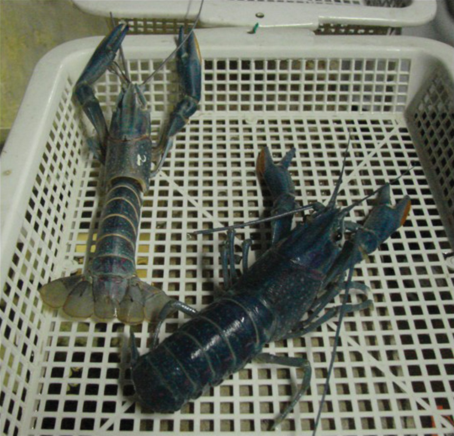
Adult specimen of the freshwater red claw crayfish, Cherax quadricarinatus, just after molting (at left the exuviae).

The storage organs are composed of a one-layered epithelium forming tubules, which are proximally connected to the midgut and distally blind-ended. During the premolt period, calcium originating from the present cuticle is transported in an ionized form and is precipitated in the PC lumen within an organic matrix synthesized by the PC cells (Figure 2C). Concretions are formed by addition of successive concentric layers of organic matrix, within which calcium carbonate is precipitated as the amorphous polymorph (Raz et al. 2002). After ecdysis, calcium resorption occurs by successive generations of 1 µm diameter calcified spherules that form at the apical part of the PC epithelium and dissolve at the basal part of the extracellular PC network. The storage proceeds exponentially during a 16-day mean period for an adult specimen concomitant with the partial demineralization of the cuticle, whereas dissolution of concretions is performed in less than 48 h. The stored calcium represents 60% of the calcium necessary for the complete calcification of each new brand cuticle.

Figure 2.

Calcium storage in the semiterrestrial amphipod, Orchestia cavimana. A Adult male specimen (2-cm long) B Radiography of an adult specimen just after ecdysis (electron-dense stored calcium is well visible in 2 diverticula of the midgut; arrows) C Calcium is stored as calcareous concretions in paired posterior ceca. c: concretion, hg: hindgut, mg: midgut.

To understand the formation of biomineralized structures, the characterization of the molecular components of the organic matrix is of first interest. Biochemical and molecular biology techniques were used to characterize proteinaceous components of the matrix. One peculiar protein, named Orchestin, has been well characterized and completely sequenced. This phosphorylated calcium-binding protein is probably responsible for the precipitation of calcium carbonate within the storage organs in premolt as well as for the formation of the calcium resorption spherules in postmolt (Testenière et al. 2002, Hecker et al. 2003, 2004). Orchestin is also thought to be involved in the determination of the amorphous calcium carbonate (ACC) polymorph, in cooperation with other matrix molecules (Hecker et al. 2004).

Other interesting amphipod models are troglobites of the genus Niphargus. They are able to store calcium in posterior ceca as amorphous calcium carbonate concretions, similarly to Orchestia amphipods, but also as rhomboedric crystalline structures (probably calcitic) within the gut (Graf 1975).

In Isopoda (Malacostraca: Peracarida)

Isopods possess a particular biphasic mode of molting: they shed first the posterior half of their cuticle, then the anterior part (Messner 1965, Steel 1993, Ziegler 1994). During the premolt period, oniscid isopods elaborate calcified deposits in the four anterior sternites between the cuticle and the hypodermis. The formation of such sternal plates has been particularly well studied in the woodlouse, Porcellio scaber (Ziegler 1994, 1997, Fabritius and Ziegler 2003, Ziegler et al. 2005). Some results were also obtained from isopods of the genus Oniscus (Steel 1993), Ligidium (Ziegler and Miller 1997, Glötzner and Ziegler 2000) and Ligia (Numanoi 1934, Ziegler and Miller 1997, Glötzner and Ziegler 2000, Štrus and Blejec 2001, Ziegler et al. 2007).

The calcified storage structures are composed of amorphous hydrated calcium carbonate precipitated within an organic matrix of spherules (Ziegler 1994, Ziegler and Miller 1997, Becker et al. 2003, Ziegler et al. 2005).

The formation and resorption of these sternal plates seem closely related to the biphasic molting cycle of these crustaceans. The calcium deposits, fully developed before ecdysis of the posterior part, are completely resorbed before ecdysis of the anterior part. It has been suggested that the calcium, stored as calcospherules in the edysial space, is used to calcify the posterior cuticle (Steel 1993, Štrus and Compère 1996, Ziegler and Merz 1999, Ziegler and Scholz 1997). After ecdysis of the anterior cuticle, the stored calcium is resorbed by the epidermis and transported until the cuticle through the haemolymph in a ionic form (Ziegler and Scholz 1997, Ziegler et al. 2005, 2007).

In Decapoda (Malacostraca: Eucarida)

The order Decapoda represents the largest group of crustaceans living on land as well as in water, and storage strategies are well developed and very diversified in decapods.

For calcifying the cuticle, calcium ions are translocated from an endogenous or exogenous source through the haemolymph. In some species this medium is used as a storage site. In the freshwater/land crab, Holthuisana transversa, the haemolymph has been observed to contain small-size calcified granules representing a way of transporting a great amount of calcium in a short period while avoiding toxification (Sparkes and Greenaway 1984).

Hepatopancreas is also used by some crabs like Cancer pagurus, Carcinus maenas, Callinectes sapidus and Paratelphusa hydrodomous as a storage organ where calcified granules of calcium phosphate have been found, also considered by some authors as playing a role in metal detoxication processes (Adiyodi 1969, Becker et al. 1974, Guary and Negrel 1981). This was also evidenced more recently in the land crab Ucides cordatus (Corrêa et al. 2002, 2009).

Finally some decapods store calcium in their cardiac stomach wall between the one-layered epithelium and a cuticle as so-called gastroliths (Travis 1960, 1963). They appear as paired semi-spherical structures in lobsters and crayfishes (Figure 3A) and as four more irregular deposits in gecarcinid land crabs. After acidic decalcification, we observed an important network of concentric and transversal micro- and nanofibers of organic matrix forming meshes of different sizes (Figure 3B and C) within which calcium carbonate is precipitated as nanospheres, as generally found in all the ACC biomineralized structures (Figure 3B).

Figure 3.

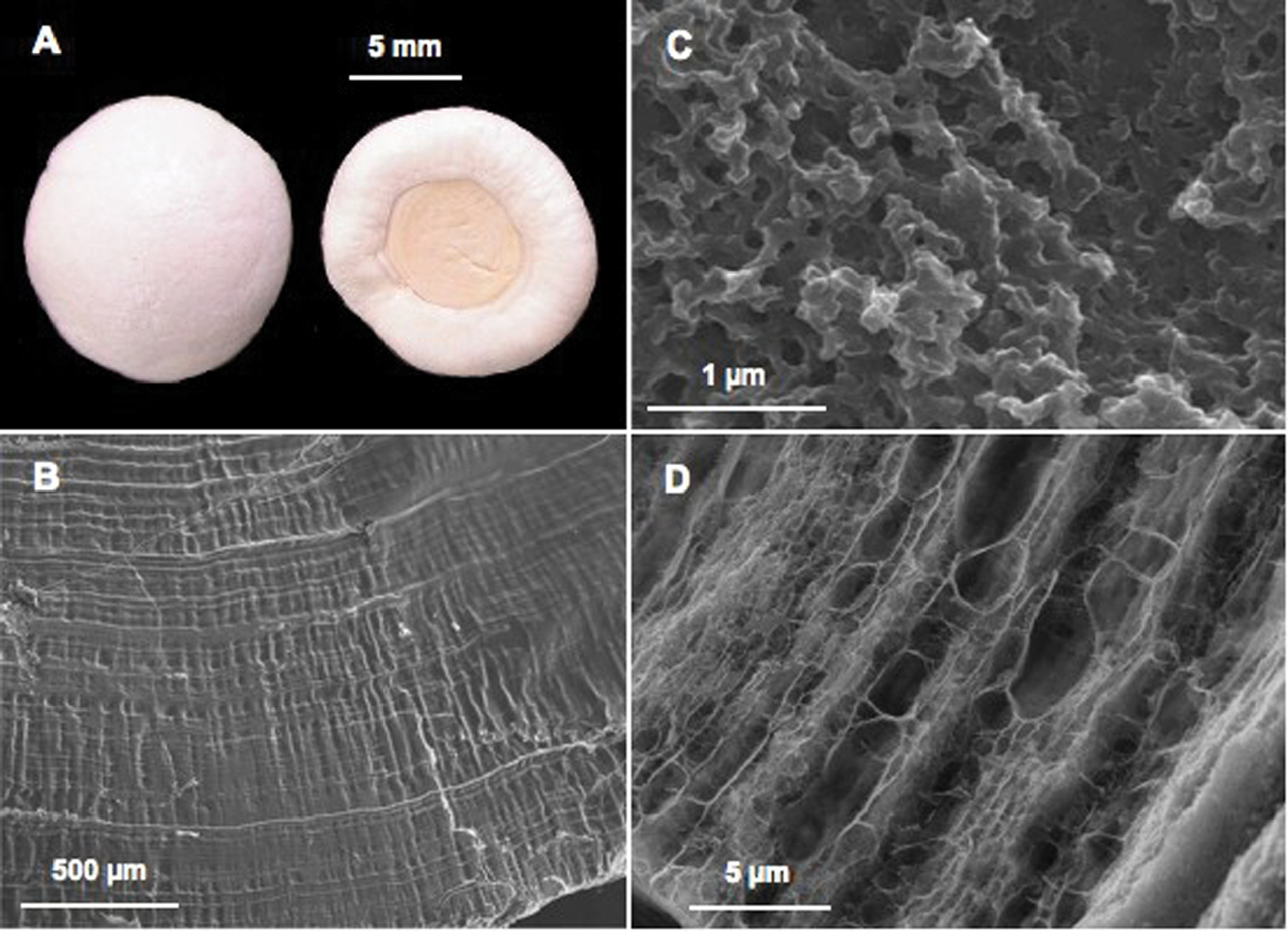
Calcium storage as gastroliths in decapods. A Pair of gastroliths from the crayfish Cherax quadricarinatus (Light Microscopy) B Internal striated structure visible on natural fracture after slight acetic acid decalcification (SEM) C Ultrastructure well visible after natural fracture and high magnification (SEM): the mineral is precipitated as nanospheres D Organic matrix network revealed after acetic acid decalcification (SEM).

Four organic matrix proteins from gastroliths have been well characterized and sequenced so far. The first one, named GAMP, was obtained from the crayfish Procambarus clarkii (Ishii et al. 1996, 1998). More recently GAP65, GAP10 and crustacyanin-A2 subunit were obtained from the Australian red claw crayfish, Cherax quadricarinatus (Shechter et al. 2008, Luquet et al. 2009, Glazer et al. 2010). Among these proteins, only GAP65 has been suggested to be involved in the determination and stabilization of the amorphous polymorph.

How the biogenic ACC polymorph can be stabilized in time is still an open question that received recent attention. It was previously suggested that specialized macromolecules (acidic proteins, phosphoproteins, sulfated glycoproteins) or ions such as magnesium or phosphate could contribute to this stabilization (Aizenberg et al. 1996, 2002, Raz et al. 2000, 2003, Addadi et al. 2003, Luquet and Marin 2004, Marin and Luquet 2007, Shechter et al. 2008, Bentov et al. 2010). Very recently, Sato and co-workers (2011) and Akiva-Tal and co-workers (2011), by using cucticle and/or gastrolith from Procambarus clarkii and Cherax quadricarinatus as models, respectively, presented a new insight into induction and stabilization of ACC. By using solid state NMR spectroscopy, they demonstrated the presence of phosphorylated energy-rich intermediates of the glycolytic pathway and suggested their possible involvement in these processes. ACC particles would form first due to the interaction of specialized matrix macromolecules bound to chitin in the CaCO3 precipitation process. Then phosphoenolpyruvate (PEP) and 3-phosphoglycerate (3PI) would act by binding to the surface of ACC, thus inhibiting the transformation of ACC into a crystalline polymorph (Sato et al. 2011). Citrate might be also involved in ACC stabilization by forming citrate-Ca2+-P complexes (Akiva-Tal et al. 2011). Similarly, it is to notice that NMR analyses revealed that the amorphous mineral storage structures found in the hepatopancreas of the crab, Ucides cordatus, are phosphate-rich granules containing mainly orthophosphate, but also pyrophosphate and glucose-6-phosphate, considered as possibly involved in the stabilization of the amorphous state (Corrêa et al. 2009).

Conclusion

The formation of the majority of biominerals is under biological control. The knowledge of the physical and chemical features of the matrix components (proteins, polysaccharides, proteoglycans, lipids, low-molecular weight components...) is a prerequisite to understand, at the molecular level, how a biomineralization is elaborated, how the matrix molecules are involved in the nucleation and precipitation processes, how they influence the determinism of the polymorph obtained, and finally how a demineralizing process may occur.

In consideration of their ability to cyclically elaborate and resorb biomineral composites, crustaceans appear as convenient models for such prospects. They are not only able to synthesize a calcified exoskeleton but also calcium storage structures, which differ in their morphology and in their mineral composition. The calcium storage deposits are transient reservoirs of calcium ions that must be quickly mobilizable after ecdysis, and, for this reason, the storage structures are composed mainly of ACC. The cuticle reveals also a remarkable biocomposite because of the simultaneous presence of different polymorphs of CaCO3 and ACP as well. Recent investigations suggest that if some general structural features may be common to all cuticles, it seems likely that the cuticle mineral composition, linked to the molecular content, not only could vary from one order of crustaceans to another but could also depend on the ecophysiology of each species.

To understand why the amorphous calcium carbonate state is stabilized in time whereas the same purely inorganic mineral is completely unstable, as well as how the switch of the transformation from the amorphous to crystalline phases occurs, is also of great interest in the biomaterial and nanotechnology fields (Han and Aizenberg 2008, Gower 2008, Tao et al. 2009). From studies on crustaceans models it was evidenced that magnesium and phosphate ions, proteinaceous macromolecules and low-molecular weight phosphorylated components of the organic matrix could be responsible for this stabilization. If some recent results appear meaningful in this sense, unfortunately there are still too few matrix molecules really well characterized so far in crustaceans, as in other phyla, to have a clear idea of the complete process of elaboration of a biomineralization. Nevertheless, it seems more and more evident that the stabilization of the amorphous state, similarly to other processes such as CaCO3 nucleation and precipitation and the simultaneous presence of different polymorphs of CaCO3 are multi-parameter phenomena, which result from the synergistic cooperation of several if not all the categories of matrix components above described.

Finally, by means of comparative studies performed in other calcifying phyla, crustaceans are useful to determine why and how calcification could have emerged on Earth. The sequence analysis of the matrix proteins and the genes encoding these proteins could lead to the understanding of the strategy used by evolution to built and select different mineralizing systems: convergence of different biological systems for a similar mineralizing function by exaptation of initially non-mineralizing molecules or evolution and adaptative divergence from an ancestral biomineral system still undeciphered?

Acknowledgements

The author is greatly indebted to Prof. François Graf for the photos of Orchestia cavimana and for many fruitful discussions on crustaceans and biomineralizations. The author also thanks Andreas Ziegler, Nada Žnidaršič and the editorial committee for valuable comments.

References
Addadi L, Raz S, Weiner S (2003) Taking advantage of disorder: amorphous calcium carbonate and its role in biomineralization. Advanced Materials 15: 959-970.
Adiyodi RG (1969) Calcium cycle and the hepatopancreas in the freshwater crab Paratelphusa. Journal of the Kerala Academy of Biology 1: 20-28.
Aizenberg J, Lambert G, Addadi L, Weiner S (1996) Stabilization of amorphous calcium carbonate by specialized macromolecules in biological and synthetic precipitates. Advanced Materials 8: 222-226.
Aizenberg J, Lambert G, Weiner S, Addadi L (2002) Factors involved in the formation of amorphous and crystalline calcium carbonate: A study of an ascidian skeleton. Journal of theAmerican Chemical Society 124: 32-39.
Akiva-Tal A, Kabaya S, Balazs YS, Glazer L, Berman A, Sagi A, Schmidt A (2011) In situ molecular NMR picture of bioavailable calcium stabilized as amorphous CaCO3 biomineral in crayfish gastroliths. Proceedings of the National Academy of Sciences USA 108: 14763-14768.
Al-Sawalmih A, Li C, Siegel S, Fabritius H, Yi S, Raabe D, Fratzl P, Paris O (2008) Microtexture and chitin/calcite orientation relationship in the mineralized exoskeleton of the American lobster. Advanced Functional Materials 18: 3307-3314.
Al-Sawalmih A, Li C, Siegel S, Fratzl P, Paris O (2009) On the stability of amorphous minerals in lobster cuticle. Advanced Materials 21: 391-400.
Andersen SO (1999) Exoskeletal proteins from the crab, Cancer pagurus. Comparative Biochemistry and Physiology A 123: 203-211.
Becker GL, Chen CH, Greenawalt JW, Lenhinger AL (1974) Calcium phosphate granules in the hepatopancreas of the blue crab Callinectes sapidus. The Journal of Cell Biology 61: 316-326.
Becker A, Bismayer U, Epple M, Fabritius H, Hasse B, Shi J, Ziegler A (2003) Structural characterization of amorphous calcium carbonate (ACC) in sternal deposits of the crustacean Porcellio scaber. Dalton Transactions: 551–555.
Becker A, Ziegler A, Epple M (2005) The mineral phase in the cuticles of two species of Crustacea consists of magnesium calcite, amorphous calcium carbonate and amorphous calcium phosphate. Dalton Transactions: 1814–1820.
Bentov S, Weil S, Glazer L, Sagi A, Berman A (2010) Stabilization of amorphous calcium carbonate by phosphate rich organic matrix proteins an by single phosphoamino acids. Journal of Structural Biology 171: 207-215.
Bouligand Y (1972) Twisted fibrous arrangement in biological material and cholestric mesophases. Tissue & Cell 4: 189-217.
Carlisle EM (1970) Silicon: a possible factor in bone calcification. Science 167: 179-280.
Chen JY, Vannier J, Huang DY (2001) The origin of crustaceans: new evidence from the Early Cambrian of China. Proceedings of the Royal Society of London B 268: 2181-2187.
Coblentz FE, Towle DW, Shafer TH (2006) Comparative Biochemistry and Physiology D 1: 200-208.
Compère P, Morgan JA, Winters C, Goffinet G (1992) X-ray microanalytical and cytochemical study of the mineralization process in the shore crab cuticle. Micron and Microscopia Acta 23: 355-356.
Compère P, Morgan JA, Goffinet G (1993) Ultrastructural location of calcium and magnesium during mineralisation of the cuticle of the shore crab, as determined by the K-pyroantimonate method and X-ray microanalysis. Cell and Tissue Research 274: 567-577.
Corrêa JD, Farina M, Allodi S (2002) Cytoarchitectural features of Ucides cordatus (Crustacea Decapoda) hepatopancreas: structure and elemental composition of electron-dense granules. Tissue & Cell 34: 315-325.
Corrêa JD, Bruno MI, Allodi S, Farina M (2009) Effects of concentration of H+ on amorphous mineral granules: Structural stability and elemental mobilization. Journal of Structural Biology 106: 59-66.
Dillaman R, Hequembourg S, Gay M (2005) Early pattern of calcification in the dorsal carapace of the blue crab, Callinectes sapidus. Journal of Morphology 263: 356-374.
Endo H, Persson P, Watanabe T (2000) Molecular cloning of the crustacean DD4 cDNA encoding a Ca2+-binding protein. Biochemical and Biophysical Research Communications 276: 286-291.
Endo H, Takagi Y, Ozaki N, Kogure T, Watanabe T (2004) A crustacean Ca2+-binding protein with a glutamate-rich sequence promotes CaCO3 crystallization. Biochemical Journal 384: 159-167.
Fabritius H, Ziegler A (2003) Analysis of CaCO3 deposits formation and degradation during the molt cycle of the terrestrial isopod Porcellio scaber (Crustacea, Isopoda). Journal of Structural Biology 142: 281-291.
Fabritius H, Sachs C, Romano Triguero P, Raabe D (2009) Influence of structural principles on the mechanics of a biological fiber-based composite material with hierarchical organization: the exoskeleton of the lobster Homarus americanus. Advanced Materials 21: 391-400.
Faircloth LM, Shafer TH (2007) Differential expression of eight transcripts and their roles in the cuticle of the blue crab, Callinectes sapidus. Comparative Biochemistry and Physiology B 146: 370-383.
Fingerman M, Naghabushanam R, Sarojini R (1993) Vertebrate-type hormones in Crustaceans: localization, identification and functional significance. Zoological Science 10: 13-29.
Giraud-Guille MM (1984a) Calcification initiation sites in the crab cuticle: the interprismatic septa. An ultrastructural cytochemical study. Cell and Tissue Research 236: 413-420.
Giraud-Guille MM (1984b) Fine structure of the chitin–protein system in the crab cuticle. Tissue & Cell 16: 75-92.
Giraud-Guille MM, Belamie E, Mosser G (2004) Organic and mineral network in carapaces, bones and biomimetic materials. Comptes Rendus Palevol 3: 503-513.
Glazer L, Shechter A, Tom M, Yudkovski Y, Well S, Aflalo ED, Pamuru RR, Khalaila I, Bentov S, Berman A, Sagi A (2010) A protein involved in the assembly of an extracellular calcium storage matrix. Journal of Biological Chemistry 285: 12831-12839.
Glötzner J, Ziegler A (2000) Morphometric analysis of the calcium-transporting sternal epithelial cells of the terrestrial isopods Ligia oceanica, Ligidium hypnorum and Porcellio scaber during molting. Arthropod Structure & Development 29: 241-257.
Gower LB (2008) Biomimetic model systems for investigating the amorphous precursor pathway and its role in biomineralization. Chemical Review 108: 4551-4627.
Graf F (1975) Chronologie du développement et évolution du stockage de calcium et des cellules à urates chez Niphargus schellenbergi Karaman. International Journal of Speleology 7: 247-272.
Graf F (1978) Les sources de calcium pour les crustacés venant de muer. Archives de Zoologie Expérimentale et Générale 119: 143-161.
Graf F, Meyran JC (1983) Premolt calcium secretion in the midgut posterior cæca of the crustacean Orchestia: ultrastructure of the epithelium. Journal of Morphology 177: 1-23.
Graf F, Meyran JC (1985) Calcium reabsorption in the posterior cæca of the midgut in a terrestrial crustacean, Orchestia cavimana. Cell and Tissue Research 242: 83-95.
Greenaway P (1985) Calcium balance and molting in the Crustacea. Biological Review 60: 425-454.
Guary JC, Négrel R (1981) Calcium phosphate granules: a trap for transuranic and iron in crab hepatopancreas. Comparative Biochemistry and Physiology A 68: 423-427.
Han TYJ, Aizenberg J (2008) Calcium Carbonate Storage in Amorphous Form and Its Template-Induced Crystallization. Chemistry of Materials 20: 1064-1068.
Hecker A, Testenière O, Marin F, Luquet G (2003) Phosphorylation of serine residues is fundamental for the calcium binding ability of Orchestin, a soluble matrix protein from crustacean calcium storage structures. FEBS Letters 535: 49-54.
Hecker A, Quennedey B, Testenière O, Quennedey A, Graf F, Luquet G (2004) Orchestin, a calcium-binding phosphoprotein, is a component of two successive transitory calcified biomineralizations elaborated by a terrestrial crustacean. Journal of Structural Biology 146: 310-324.
Hild S, Marti O, Ziegler A (2008) Spatial distribution of calcite and amorphous calcium carbonate in the cuticle of the terrestrial crustaceans Porcellio scaber and Armadillidium vulgare. Journal of Structural Biology 163: 100-108.
Hild S, Neues F, Znidarsic N, Štrus J, Epple M, Marti O, Ziegler A (2009) Ultrastructure and mineral distribution in the tergal cuticle of the terrestrial isopod Titanethes albus. Adaptations to a karst cave biotope. Journal of Structural Biology 168: 426-436.
Inoue H, Ozaki N, Nagasawa H (2001) Purification and structural determination of a phosphorylated peptide with anti-calcification and chitin-binding activities in the exoskeleton of the crayfish, Procambarus clarkii. Bioscience Biotechnology and Biochemistry 65: 1840-1848.
Inoue H, Ohira T, Ozaki N, Nagasawa H (2003) Cloning and expression of a cDNA encoding a matrix peptide associated with calcification in the exoskeleton of the crayfish, Comparative Biochemistry and Physiology B 136: 755–765.
Inoue H, Ohira T, Ozaki N, Nagasawa H (2004) A novel calcium-binding peptide from the cuticle of the crayfish, Procambarus clarkii. Biochemical Biophysical Research Communications 318: 649-654.
Inoue H, Ohira T, Nagasawa H (2007) Significance of the N- and C-terminal regions of CAP-1, a cuticle calcification-associated peptide from the exoskeleton of the crayfish, for calcification. Peptides 28: 566-573.
Inoue H, Yuasa-Hashimoto N, Suzuki M, Nagasawa H (2008) Structural determination and functional analysis of a soluble matrix proteins associated with calcification of the exoskeleton of the crayfish, Procambarus clarkii. Bioscience Biotechnology and Biochemistry 72: 2697-2707.
Ishii K, Yanagisawa T, Nagasawa H (1996) Characterization of a matrix protein in the gastroliths of the crayfish Procambarus clarkii. Bioscience Biotechnology and Biochemistry 60: 1479-1482.
Ishii K, Tsutsui N, Watanabe T, Yanagisawa T, Nagasawa H (1998) Solubilization and chemical characterization of an insoluble matrix protein in the gastroliths of a crayfish, Procambarus clarkii. Bioscience Biotechnology and Biochemistry 62: 291-296.
Kirschvink JL, Hagadorn JW (2000) A grand unified theory of biomineralization. In: Bäuerlein E (Ed) Biomineralization, Wiley-VCH Verlag Gmbh, Weinheim, Germany, 139–150.
Knoll AH (2004) Biomineralization and evolutionary history. In: Dove PM, DeYoreo JJ, Weiner S (Eds) Reviews in Mineralogy and Geochemistry 54: 329-356.
Kragh M, Mølbak L, Andersen SO (1997) Cuticular proteins from the lobster, Homarus americanus. Comparative Biochemistry and Physiology B 118: 147-54.
Kuballa AV, Merritt DJ, Elizur A (2007) Gene expression profiling of cuticular proteins across the moult cycle of the crab Portunus pelagicus. BMC Biology 5: 45-70. doi: 10.1186/1741-7007-5-45
Kuballa AV, Elizur A (2008) Differential expression profiling of components associated with exoskeletal hardening in crustaceans. BMC Genomics 9: 575-588. doi: 10.1186/1471-2164-9-575
Kuballa AV, Holton TA, Paterson B, Elizur A (2011) Molt cycle specific differential gene expression profiling of the crab Portunus pelagicus. BMC Genomics 12: 147-164. doi: 10.1186/1471-2164-12-147
Landis WJ, Lee DD, Brenna JT, Chandra S, Morrison GH (1986) Detection and localization of silicon and associated elements in vertebrate bone tissue by imaging ion microscopy. Calcified Tissue International 38: 52-59.
Lowenstam HA, Weiner S (1989) On biomineralization, Oxford University Press, NewYork, USA, 324 pp.
Lowenstam HA (1981) Minerals formed by organisms. Science 211: 1126-1131.
Luquet G, Marin F (2004) Biomineralizations in crustaceans: storage strategies. Comptes Rendus Palevol 3: 515-534.
Luquet G, Le Roy N, Zanella-Cléon I, Becchi M, Bucarey S, Fernandez MS, Arias JL, Guichard N, Marie B, Marin F (2009) Characterization of Crustacyanin-A2 Subunit as a component of the organic matrix of gastroliths from the crayfish Cherax quadricarinatus. In: Kisalius D, Estroff L, Landis W, Zavattieri P, Gupta HS (Eds) Structure-Property Relationships in Biomineralized and Biomimetic Composites, Warrendale, Material Research Society Symposium Proceedings 1187: 69-75.
Magkrioti CK, Spyropoulos IC, Iconomidou VA, Willis JH, Hamodrakas SJ (2004) CuticleDB: a relational database of Arthropod cuticular proteins. BMC Bioinformatics 5: 138. doi: 10.1186/1471-2105-5-138
Mann S (1983) Mineralization in biological systems. Structural Bonding 54: 125-174.
Marin F, Luquet G (2007) Unusually acidic proteins in biomineralization. In: Bäuerlein E, Behrens P, Epple M (Eds). Handbook of Biomineralization, Vol. 1: The Biology of Biominerals Structure Formation. Wiley-VCH: 273-290.
Matsko NB, Žnidaršič N, Letofsky-Papst L, Dittrich M, Grogger W, Štrus J, Hofer F (2011) Silicon: The key element in early stages of biocalcification. Journal of Structural Biology 174: 180-186.
McWhinnie MA, Cahoon MO, Johanneck R (1969) Hormonal effects on calcium metabolism in Crustacea. American Zoologist 9: 841-855.
Messner B (1965) Ein morphologish-histologisher Beitrag zur Häutung von Porcellio scaber Latr. und Oniscus asellus L. (Isopoda terrestria). Crustaceana 9: 285-301.
Meyran JC, Graf F, Nicaise G (1984) Calcium pathway through a mineralizing epithelium in the crustacean Orchestia in premolt: ultrastructural cytochemistry and X-ray microanalysis. Tissue & Cell 16: 269-286.
Meyran JC, Graf F, Nicaise G (1986) Pulse discharge of calcium through a demineralizing epithelium in the crustacean Orchestia: ultrastructural cytochemistry and X-ray microanalysis. Tissue & Cell 18: 267-283.
Neues F, Ziegler A, Epple M (2007) The composition of mineralized cuticle in marine and terrestrial isopods: a comparative study. Crystal Engineering Communications 9: 1245-1251.
Neues F, Hild S, Epple M, Marti O, Ziegler A (2011) Amorphous and crystalline calcium carbonate distribution in the tergite cuticle of moulting Porcellio scaber (Isopoda, Crustacea). Journal of Structural Biology 175: 10-20.
Nikolov S, Petrov M, Lymperakis L, Friak M, Sachs C, Fabritius H, Raabe D, Neugebauer J (2010) Revealing the design principles of high-performance biological composites using ab initio and multiscale simulations: the example of lobster cuticle. Advanced Materials 22: 519-526.
Nikolov S, Fabritius H, Petrov M, Friak M, Lymperakis L, Sachs C, Raabe D, Neugebauer J (2011) Robustness and optimal use of design principles of arthropod exoskeletons studied by ab initio-based multiscale simulations. Journal of The Mechanical Behavior of Biomedical Materials 4: 129-145.
Nousiainen M, Rafn K, Skou L, Roepstorff P, Andersen SO (1998) Characterization of exoskeletal proteins from the american lobster, Homarus americanus. Comparative Biochemistry and Physiology B 119: 189-199.
Numanoi H (1934) Calcium contents of the carapace and other organs of Ligia exotica during non-molting and molting phases. Journal of the Faculty of Science of the Imperial University of Tokyo, Zoology 3: 359-364.
Pouget E, Boman P, Goos J, Frederik P, de With G, Sommerdijk N (2009) The initial stages of template-controlled CaCO3 formation revealed by cryo-TEM. Science 323: 1455-1458.
Price JB, Holdich DM (1980a) The formation of the epicuticle and associated structures in Oniscus asellus (Crustacea, Isopoda). Zoomorphology 94: 321-332.
Price JB, Holdich DM (1980b) An ultrastructural study of the integument during the moult cycle of the woodlouse, Oniscus asellus (Crustacea, Isopoda). Zoomorphology 95: 250-263.
Raabe D, Al-Sawalmih A, Yi S, Fabritius H (2008) Preferred crystallographic texture of a-chitin as a microscopic and macroscopic design principle of the exoskeleton of the lobster Homarus americanus. Acta Biomaterialia 3: 882-895.
Raz S, Weiner S, Addadi L (2000) Formation of high-magnesian calcites via an amorphous precursor phase: possible biological implications. Advanced Materials 12: 38-42.
Raz S, Testenière O, Hecker A, Weiner S, Luquet G (2002) Amorphous calcium carbonate is the main component of the calcium storage structures of the crustacean Orchestia cavimana. Biological Bulletin 203: 269-274.
Raz S, Hamilton PC, Wilt FH, Weiner S, Addadi L (2003) The transient phase of amorphous calcium carbonate in sea urchin larval spicules: The involvement of proteins and magnesium ions in its formation and stabilization. Advanced Functional Materials 13: 480-486.
Rebers JE, Willis JH (2001) A conserved domain in arthropod cuticular proteins binds chitin. Insect Biochemistry and Molecular Biology 31: 1083-1093.
Roer R (1980) Mechanisms of resorption and deposition of calcium in the carapace of the crabCarcinus maenas. Journal of Experimental Biology 8: 205-218.
Roer R, Dillaman R (1984) The structure and calcification of the crustacean cuticle. American Zoologist 24: 893-909.
Romano P, Fabritius H, Raabe D (2007) The exoskeleton of the lobster, Homarus americanus, as an example of a smart anisotropic biological material. Acta Biomaterialia 3: 301-309.
Sachs C, Fabritius H, Raabe D (2006) Experimental investigation of the elastic-plastic deformation of mineralized lobster cuticle by digital image correlation. Journal of Structural Biology 155: 409-425.
Sato A, Nagasaka S, Fuhirata K, Nagata S, Arai S, Saruwatari K, Kogure T, Sakuda S, Nagasawa H (2011) Glycolytic intermediates induce amorphous calcium carbonate formation in crustaceans. Nature Chemical Biology 7: 197-199.
Seidl B, Huemer K, Neues F, Hild S, Epple M, Ziegler A (2011) Ultrastructure and mineral distribution in the tergite cuticle of the beach isopod Tylos europeus Arcangeli, 1938. Journal of Structural Biology 174: 512-526.
Shafer TH, McCartney MA, Faircloth LM (2006) Identifying exoskeleton proteins in the blue crab from an expressed sequence tag (EST) library. Integrative and Comparative Biology 46: 978-990.
Shechter A, Glazer L, Cheled S, Mor E, Weil S, Berman A, Bentov S, Aflalo ED, Khalaila I, Sagi A (2008) A gastrolith protein serving a dual role in the formation of an amorphous mineral containing extracellular matrix. Proceedings of the National Academy of Sciences USA 105: 7129-7134.
Simkiss K, Wilbur KM (1989) Biomineralization: Cell Biology and Mineral Deposition, Academic Press, San Diego, USA, 337 pp.
Sparkes S, Greenaway P (1984) The hemolymph as a storage site for cuticular ions during premolt in the freshwater/land crab Holthuisana transversa. Journal of Experimental Biology 113: 43-54.
Steel CGH (1993) Storage and translocation of integumentary calcium during the molt cycle of the terrestrial isopod Oniscus asellus (L.). Canadian Journal of Zoology 71: 4-10.
Štrus J, Compère P (1996) Ultrastructural analysis of the integument during the moult cycle in Ligia italica (Crustacea, Isopoda). European Journal of Physiology 431(Suppl): R251-R252.
Štrus J, Blejec A (2001) Microscopic anatomy of the integument and digestive system during the molt cycle in Ligia italica (Oniscidea). In: Kensley B, Brusca RC (Eds) Crustacean Issues 13, “Isopod systematics and evolution”, AA Balkema Publishers, Rotterdam, 343–352.
Sugawara A, Nishimura T, Yamamoto Y, Inoue H, Nagasawa H, Kato T (2006) Self-Organization of Oriented Calcium Carbonate/Polymer Composites: Effects of a Matrix Peptide Isolated from the Exoskeleton of a Crayfish. Angewandte Chemie International Edition 45: 1-5.
Tao J, Zhou D, Zhang Z, Xu X, Tang R (2009) Magnesium-aspartate-based crystallization switch inspired from shell molt of crustacean. Proceedings of the National Academy of Sciences USA 106: 22096-22101.
Testenière O, Hecker A, Le Gurun S, Quennedey B, Graf F, Luquet G (2002) Characterization and spatiotemporal expression of orchestin, a gene encoding an ecdysone-inducible protein from a crustacean organic matrix. Biochemical Journal 361: 327-335.
Travis DF (1960) The deposition of skeletal structures in the crustacea. I. The histology of the gastrolith skeletal tissue complex and the gastrolith in the crayfish, Orconectes (Cambarus) virilis Hagen - Decapoda. Biological Bulletin 18: 137-149.
Travis DF (1963) Structural features of mineralization from tissue to macromolecular levels of organization in the decapod Crustacea. Annals of the New York Academy of Sciences 109: 177-245.
Wood S, Russell JD (1987) On the nature of the calcium carbonate in the exoskeleton of the woodlouse Oniscus asellus L. (Isopoda, Oniscoidea). Crustaceana 53: 49-53.
Wynn A, Shafer TH (2005) Four differentially expressed cDNAs in Callinectes sapidus containing the Rebers-Riddiford consensus sequence. Comparative Biochemistry and Physiology B 141: 294-306.
Yamamoto Y, Nishimura T, Sugawara A, Inoue H, Nagasawa H, Kato T (2008) Effects of peptides on CaCO3 crystallization: mineralization properties of an acidic peptide isolated from the exoskeleton of crayfish and its derivatives. Crystal Growth & Design 8: 4062-4065.
Ziegler A (1994) Ultrastructure and electron spectroscopic diffraction analysis of the sternal calcium deposits of Porcellio scaber Latr. (Isopoda, Crustacea). Journal of Structural Biology 112: 110-116.
Ziegler A (1997) Ultrastructure changes of the anterior and posterior sternal integument of the terrestrial isopod Porcellio scaber (Crustacea) during the molt cycle. Tissue & Cell 29: 63-76.
Ziegler A, Miller B (1997) Ultrastructure of CaCO3 deposits of terrestrial Isopods (Crustacea, Oniscidea). Zoomorphology 117: 181-187.
Ziegler A, Scholz FHE (1997) The ionic hemolymph composition of the terrestrial isopod Porcellio scaber Latr. during molt. Journal of Comparative Physiology 167: 536-542.
Ziegler A, Merz E (1999) Membrane particle distribution in the sternal epithelia of the terrestrial isopod Porcellio scaber Latr. (Crustacea, Oniscidea) during CaCO3 deposit formation and resorption, a freeze-etch analysis. Journal of Structural Biology 127: 263-278.
Ziegler A, Fabritius H, Hagedorn M (2005) Microscopical and functional aspects of calcium-transport and deposition in terrestrial isopods. Micron 36: 137-153.
Ziegler A, Hagedorn M, Ahearn A, Carefoot TH (2007) Calcium translocation during the molting cycle of the semi-terrestrial isopod Ligia hawaiiensis. Journal of Comparative Physiology 177: 99-108.