


(C) 2010 Xin Xu. This is an open access article distributed under the terms of the Creative Commons Attribution License, which permits unrestricted use, distribution, and reproduction in any medium, provided the original author and source are credited.
For reference, use of the paginated PDF or printed version of this article is recommended.
The taxonomic status of the genus Ambengana Millidge & Russell-Smith, 1992, is revised on the basis of its original description, illustrations and re-examination of the type species. A new synonymy is proposed: Ambengana complexipalpis Millidge & Russell-Smith, 1992 (the type species of Ambengana) syn. n. with Neriene birmanica (Thorell, 1887). Therefore, the genus Ambengana Millidge & Russell-Smith, 1992 syn. n. is synonymized with Neriene Blackwall, 1833. A morphological re-description, diagnosis and comparative illustrations are provided for Neriene birmanica as well.
Taxonomy, Linyphiidae, Neriene, Ambengana, new synonymy
The monotypic genus Ambengana Millidge & Russell-Smith, 1992, was erected for Ambengana complexipalpis Millidge & Russell-Smith, 1992, from Bali (
Neriene birmanica was first described as Linyphia birmanica from the female (
Specimens were examined with an Olympus SZX16 stereomicroscope; details were studied with an Olympus BX51 compound microscope. Male palps and female epigyne were examined and illustrated after being dissected from the spider bodies. Spermathecae were cleared in boiling KOH solution to dissolve soft tissues, and the embolic divisions of male palps were excised by breaking the column (the membranous connection between the suprategulum and the radix). Photos were made with Cannon G10 digital camera (14.7 megapixels) mounted on an Olympus SZX16 dissecting microscope. The digital images depicting the general appearance and genital morphology are a composite of multiple images taken at different focal lengths along the Z axis and assembled using the software package Helifocus 3.10. Left structures (e.g., palps, legs, etc.) are depicted unless otherwise stated. Most hairs and macrosetae are usually not depicted in the final palp and epigynum images.
All measurements were obtained using an Olympus SZX16
stereomicroscope and are given in millimeters. Eye diameters are taken
at the widest point. The total body length does not include the length
of the chelicerae or spinnerets. The leg measurements are given in the
following sequence: total (femur, patella +tibia, metatarsus,
tarsus). The terminology used in text and figure legends follows
The following abbreviations are used in the text and figures. Male palp: ALP- anterior projection of lamella; DLP- dorsal projection of lamella; E- Embolus; MM- median membrane; MA-median apophysis; L- lamella; LLP- lateral projection of lamella; P- paracymbium; PLP- posterior projection of lamella; ST- subtegulum; T- tegulum; TA- terminal apophysis. Epigynum: FG- fertilization groove; S- spermatheca; SC- “scape”; SG- spiral groove; TP- turning point. Somatic characters: AER- anterior eye row; ALE- anterior lateral eye; AME- anterior median eye; AME-ALE- distance between AME and ALE; AME-AME- distance between AMEs; AMEd- diameter of AME; PER- posterior eye row; PLE- posterior lateral eye; PME- posterior median eye; PMEd- diameter of PME; PME-PLE- distance between PME and PLE; PME-PME- distance between PMEs.
All the specimens examined are deposited in the College of Life Sciences, Hubei University, China.
TaxonomyLinyphia clathrata Sundevall, 1830.
Figs 1–12
CHINA: 1 ♂, 3 ♀, Sichuan Province, Panzhihua City, Miyi County, 10.07.1981 Gao J.C.; 1 ♂, 1 ♀, same locality, Chen X.E.; 2 ♂, 3 ♀, Yunnan Province, Dehong DaiJingpo Autonomous Prefecture, Ruili City, 16.09.2000, Chen, W. H. and Liu, F. X.; 2 ♂, 6 ♀, Yunnan Province, Lincang City, Zhenkang County, Nansan Town, 14.09.2000, Chen, W. H. and Liu, F. X.; 1 ♀, Yunnan Province, Honghe Hani and Yi Prefecture, Luchun County, 2.09.2000, Chen, W. H. and Liu, F. X.; 3 ♂, 7 ♀, Yunnan Province, Xishuangbanna Dai Autonomous Prefecture, Menghai County, Daluo Town, 9.09.2000, Chen, W. H. and Liu, F. X.; 1 ♂, same province and prefercture, Menghai County, 10.09.2000, Chen, W. H. and Liu, F. X.; 1 ♀, Yunnan Province, Honghe Hani and Yi Prefecture, Jinping Miao, Yao and Dai Autonomous County, 30.08.2000, Chen, W. H. and Liu, F. X.; 1 ♀, Yunnan Province, Pu'er Prefecture, Jiangcheng Hani and Yi Autonomous County, 25.09.2000, Chen, W. H. and Liu, F. X.
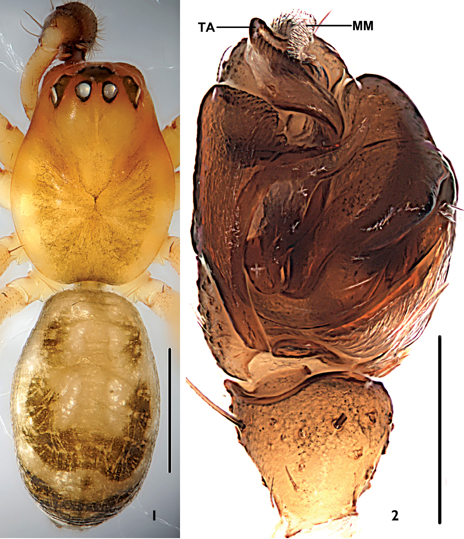
Neriene birmanica (Thorell, 1887), male from Nansan Town (Yunnan Province, China). 1 male habitus, dorsal view 2 left palp, ventral view. Scale bars =1 mm (1), scale = 0.2 mm (2).
Tibia not fusiform, with few spines; paracymbium with narrow distal arm tapering to a sharp tip; hook-shaped tip of distal part of median apophysis curved in ventral direction; transversal and terminal sclerites never present; lateral depressions of the epigyne small and superficial, this species belongs to the peltata-species group (van Helsdingen 1969). It can be distinguished from other members of the peltata-group species by the tiny paracymbium (Fig. 4), the sword-shaped embolic tip (Figs 4, 6), the broad and short terminal apophysis with about one coil (Figs 2, 5), the translucent spot at either side of the uniquely trapeziform atrium opening, the superficially depressed area at either side next to lateral translucent spots (Fig. 9), and the scape forming a rounded mesal projection and spiral grooves of about two coils in the female (Figs 10, 11).
Male: Total length: 2.78. Carapace: 1.22 long, 0.94 wide. Abdomen: 1.54 long, 0.98 wide. Carapace brown, unmodified. Eyes subequal. AER recurved, AME-AME shorter than AMEd, AME-ALE slightly longer than AMEd; PER straight, PME-PME about PMEd, PME-PLE slightly longer than PMEd; ALE and PLE juxtaposed. Chelicerae brown, stridulatory ridges absent, promargin of fang groove with three teeth, median tooth largest;, retromargin with three small teeth, first bigger than others. Lengths of legs: I 5.36 (1.47+1.58+1.52+0.79), II 4.90 (1.33+1.43+1.38+0.76), III 4.08 (1.19+1.16+1.08+0.65), IV 4.41 (1.25+1.21+1.29+0.66). Each tibia, patella and femur with two dorsal spines. Tm I: 0.20. Tm IV absent. Abdomen cylindriform, without tubercle; colour and pattern as in Fig. 1.
Patella short, with long spine dorsally. Tibia shorter than cymbium, with several long spines on lateral and ventral surfaces, and one prodorsal, two retrodorsal trichobothria. Paracymbium tiny, long and slender, U-shaped, slightly membranous (Fig. 4). Median apophysis long, slender in lateral view, with dorsal tip hook-shaped (Figs 4, 7). Lamella well-developed, with four projections: lateral one and posterior one long, lateral one with sharp, membranous end, posterior one with blunt end; anterior one broad, blunt; dorsal one short (Fig. 3). Terminal apophysis simple, broad and short, strongly membranous from prolateral view, with about one coil (Figs 2, 5). Embolus simple, perpendicularly curved at half length, with a sword-shaped end (Figs 4, 6).
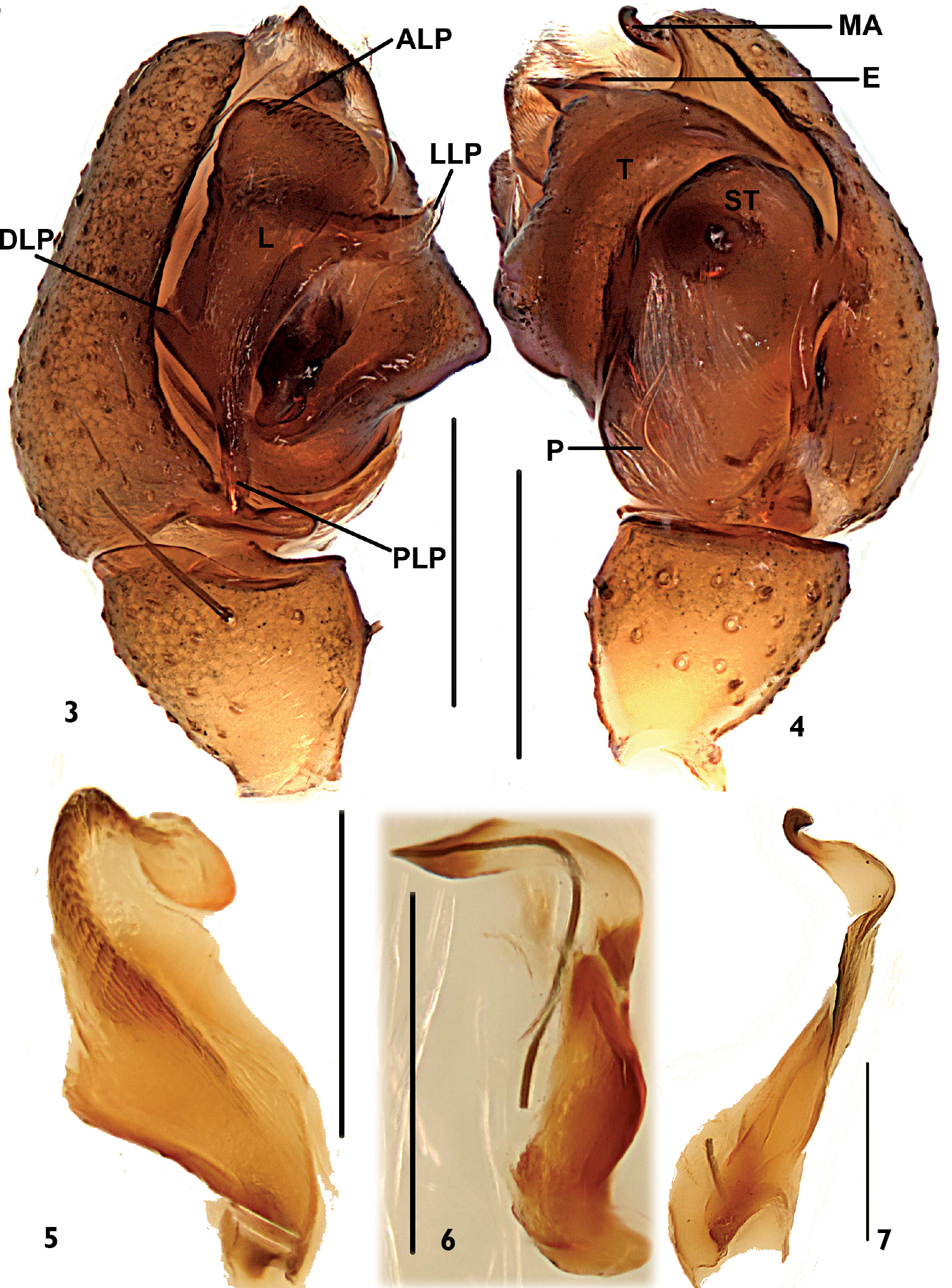
Neriene birmanica (Thorell, 1887), male from Nansan Town (Yunnan Province, China). 3 left palp, prolatral view 4 left palp, retrolateral view 5 terminal apophysis, retrolateral view 6 embolus, retrolateral view 7 median apophysis, retrolateral view. Scale bars = 0.2 mm.
Female: Total length: 2.86. Carapace: 1.14 long, 0.69 wide. Abdomen: 1.71 long, 1.30 wide. Carapace brown, unmodified. Eyes subequal. AER recurved, AME-AME shorter than AMEd, AME-ALE slightly shorter than AMEd; PER straight, PME-PME shorter than PMEd, PME-PLE about equal with PME-PME; ALE and PLE juxtaposed. Chelicerae brown, stridulatory ridges absent, promargin of fang groove with three teeth, median tooth largest; retromargin with three equal teeth. Lengths of legs: I 5.27 (1.44+1.61+1.44+0.78), II 4.60 (1.26+1.42+1.23+0.69), III 3.17 (0.93+0.94+0.79+0.51), IV 4.47 (1.37+1.27+1.21+0.62). Each tibia, patella and femur with two dorsal spines. Tm I: 0.22. Tm IV absent. Abdomen oval, without tubercle, the colour and patch see Fig. 8.
In ventral view, atrium opening small, trapeziform. Lateral depression present (Fig. 9). Scape arising from dorsal wall, short, with slightly rounded tip, and with small semi-covered depression on ventral surface (Fig. 10). Spermathecae as long as wide; spiral grooves started from entrances situated in the middle of the ventral wall of either atrium to the apical turning-points, with about two coils; fertilized ducts started from spermathecae, with (about) two coils; turning points situated laterally; spermathecae long and slender, situated laterally (Fig. 11).
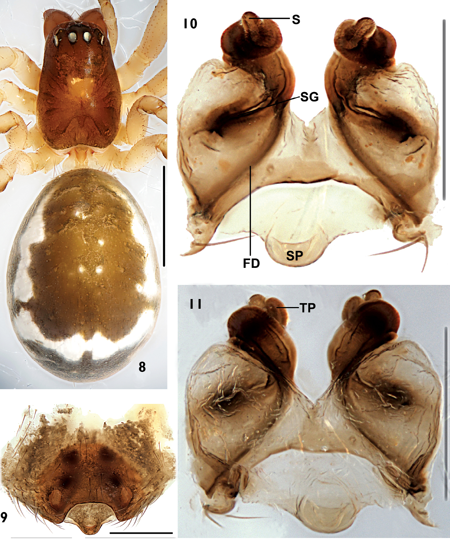
Neriene birmanica (Thorell, 1887), female from Nansan Town (Yunnan Province, China). 8 female habitus, dorsal view 9 epigynum, ventral view 10 epigynum without skin, ventral view 11 spermathecae, dorsal view. Scale = 1 mm (8), scales = 0.2 mm (9, 10, 11).
Although we didn’t examine the type speciemens of Ambengana complexipalpis,
the tiny paracymbium, the sword-shape embolic tip, broad and the
short terminal apophysis, the uniquely trapeziform atrium opening shown
in the original illustrations (
Southeast Asia (India, Kashmir, Myanmar, China, Indonesia) (Fig. 12).
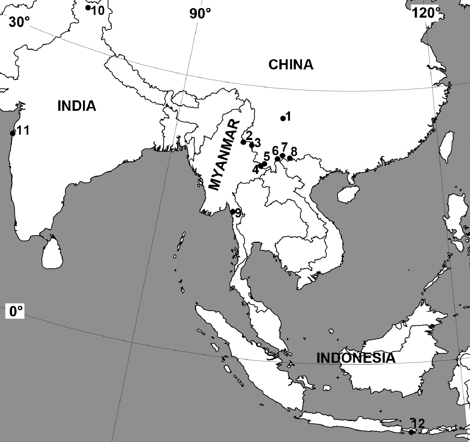
Distribution of Neriene birmanica (Thorell, 1887). Numbers correspond to the localities on the map: 1 China, Sichuan Province, Miyi County 2 China, Yunnan Province, Ruili City 3 China, Yunnan Province, Zhenkang County 4 China, Yunnan Province, Menghai County 5 China, Yunnan Province, Menghai County 6 China, Yunnan Province, Jiangcheng Hani and Yi Autonomous County 7 China, Yunnan Province, Luchun County 8 China, Yunnan Province, Jinping Miao, Yao, and Dai Autonomous County 9 Myanmar, Mawlamyine, Burma 10 Kashmir, Garhi near Jhelum 11 India, Bombay 12 Indonesia, Bali.
The manuscript benefited from the comments of Dr. Daiqin Li (National University of Singapore) and of two anonymous reviewers. The authors also thank Hao Yu (Hubei University, China) for taking the measurements of the specimens. This study was supported by the National Natural Sciences Foundation of China (NSFC-39870105/30370206), and by the Ministry of Science and Technology of the People’s Republic of China (MOST grant # 2006FY120100/2006FY110500), and by Kadoorie Farm and Botanic Garden of Hong Kong. The English of the final draft was kindly edited by Dr Robin Leech (Canada).