|
||
|
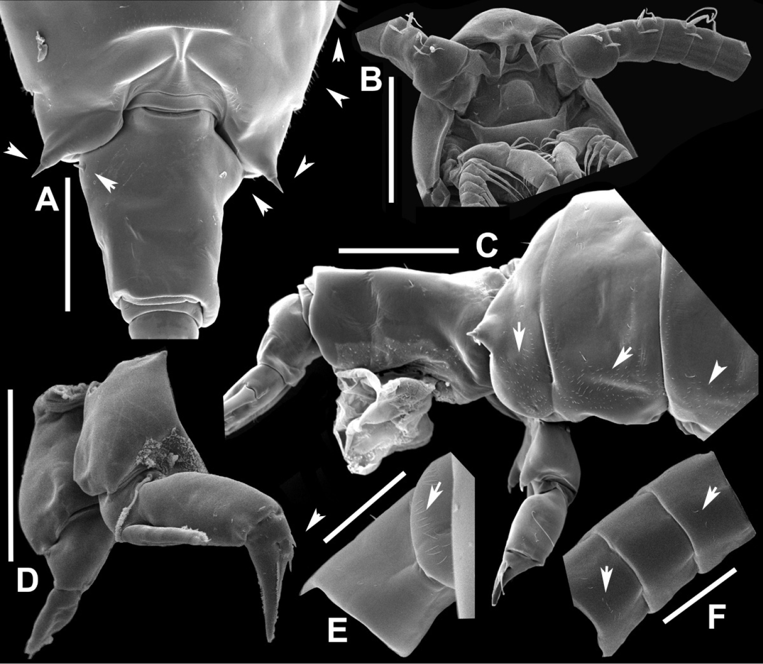
Notodiaptomus henseni female, SEM photographs. A Dorsal view of posterior part of prosome and GS (300 µm) B Ventral view of anterior end of cephalothorax, with rostrum and basal segments of antennule (150 µm) C Lateral view of posterior part of prosome, with arrows showing surface ornamentation of spinules, GS, urosome and CR (100 µm) D P5 (100 µm) E Dorsal view of segments 1 and 2 of left antennule, showing spinules (50 µm) F Dorsal view of segments 3–5 of left antennule, with some spinules (50 µm). |