|
||
|
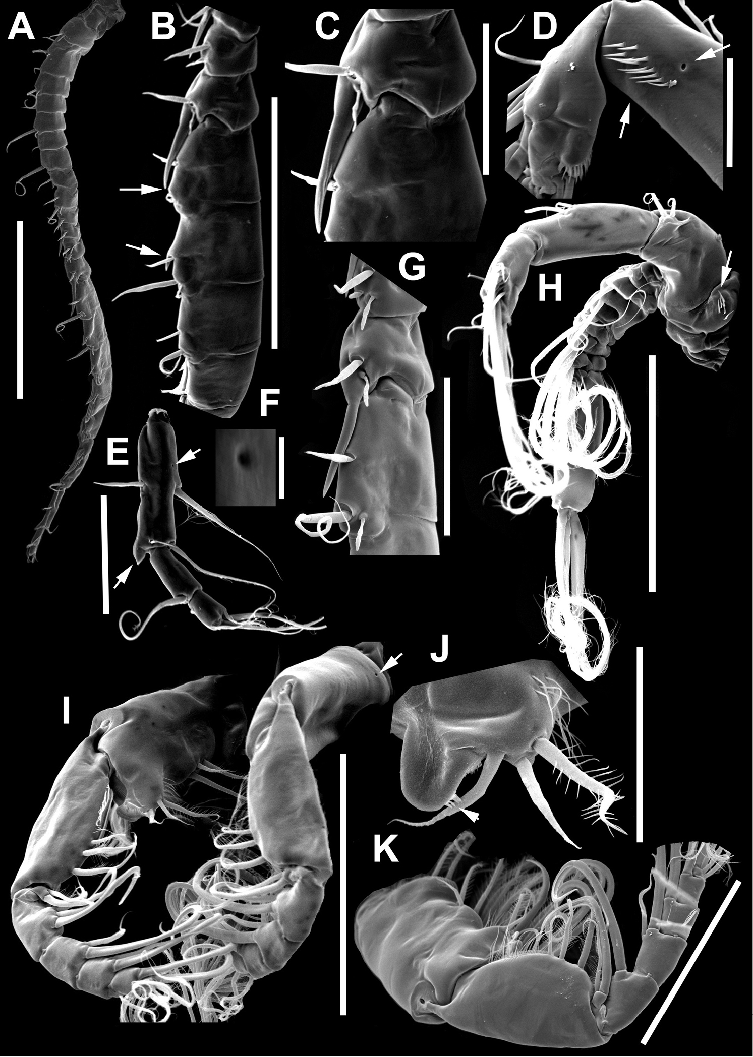
Argyrodiaptomus azevedoi male, SEM photographs. A Geniculate right antennule (500 µm) B Segments 12–17 of A1R (300 µm) C Segments 13–14 of A1R (100 µm) D Enp of A2, showing pore and spinular ornamentation (50 µm) E Segments 20–22 of A1R (100 µm) F Inset showing pore on segment 20 of A1R (5 µm) G Segments 12–16 of A1R (100 µm) H A2 (200 µm) I Maxillipeds (200 µm) J Distal endite of maxilliped (50 µm) K Maxilliped (200 µm). |