|
||
|
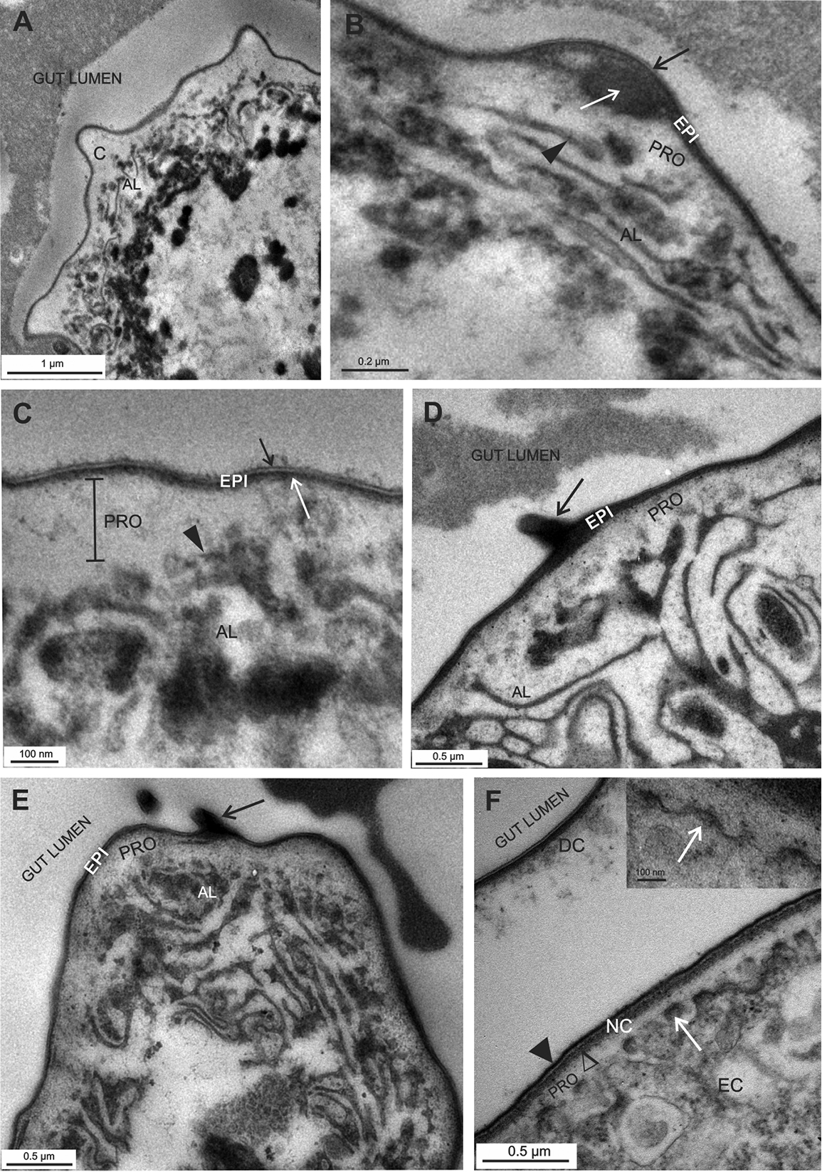
Cuticle in the hindgut of P. scaber marsupial mancae. EC - epithelial cell, PRO – procuticle, EPI – epicuticle, AL – apical labyrinth. A, B, C The hindgut cuticle (C) in early marsupial manca with the outer epicuticle and the inner procuticle. The epicuticle (EPI) consists of the outermost trilayered lamina (B, C - black →) and electron dense material underneath (C - white →). The procuticle (PRO) contains homogenous electron lucent material. Bulges of the cuticle are observed, some include electron dense material (B white →). Apical plasma membrane is intensely invaginated (B, C – ►) and forms apical labyrinth (AL) D, E The hindgut cuticle in late marsupial manca in the anterior chamber (D) and in the papillate region (E). Electron dense material is prominent under the trilayered lamina of the epicuticle. Cuticular spines are evident (black →) F Hindgut cuticle renewal in late marsupial manca - degradation and detachment of the old cuticle (DC) and formation of the new cuticle (NC) on the plasma membrane protrusions (white →). The new cuticle consists of an electron dense lamina (►), an electron dense material accumulating underneath (∆) and an inner electron lucent homogenous procuticle (PRO) F inset: Protrusions of the apical plasma membrane (white →) display electron dense tips – plaques – and are covered with an electron dense material. |