|
||
|
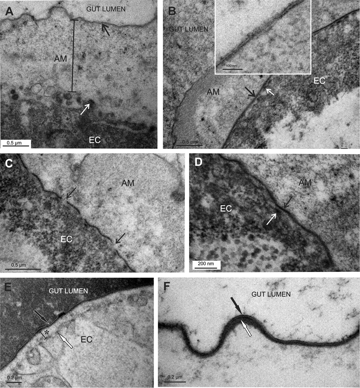
Apical matrices in the hindgut of P. scaber late embryos. EC - epithelial cell A The hindgut cells (EC) in stage 16 embryos are covered by a substantial apical matrix with intensely ruffled surface (AM). The matrix consists of an electron dense lamina (black →) and underlying more electron lucent homogenous material. The apical membrane displays irregularly arranged protrusions (white →) B, C, D In the stage 18 embryos the apical matrix of the hindgut (AM) is extensive. The surface lamina covers the matrix, which displays a distal region of medium density and a proximal lucent region. The lamina of this matrix is trilayered (B inset). A new electron dense lamina (B, D - black →) is evident above the apical membrane protrusions (B, D - white →). The new lamina is mostly continuous, though in some regions it still appears in fragments (C - black →) E, F In the prehatching embryo of stage 19 the hindgut apical matrix consists of a distal trilayered lamina (black →), an electron dense material, accumulating underneath the lamina (F - white →) and underlying lucent material (E - *). Microvilli-like protrusions of the apical plasma membrane are evident (E - white →). The gut lumen is filled with homogenous material. |