|
||
|
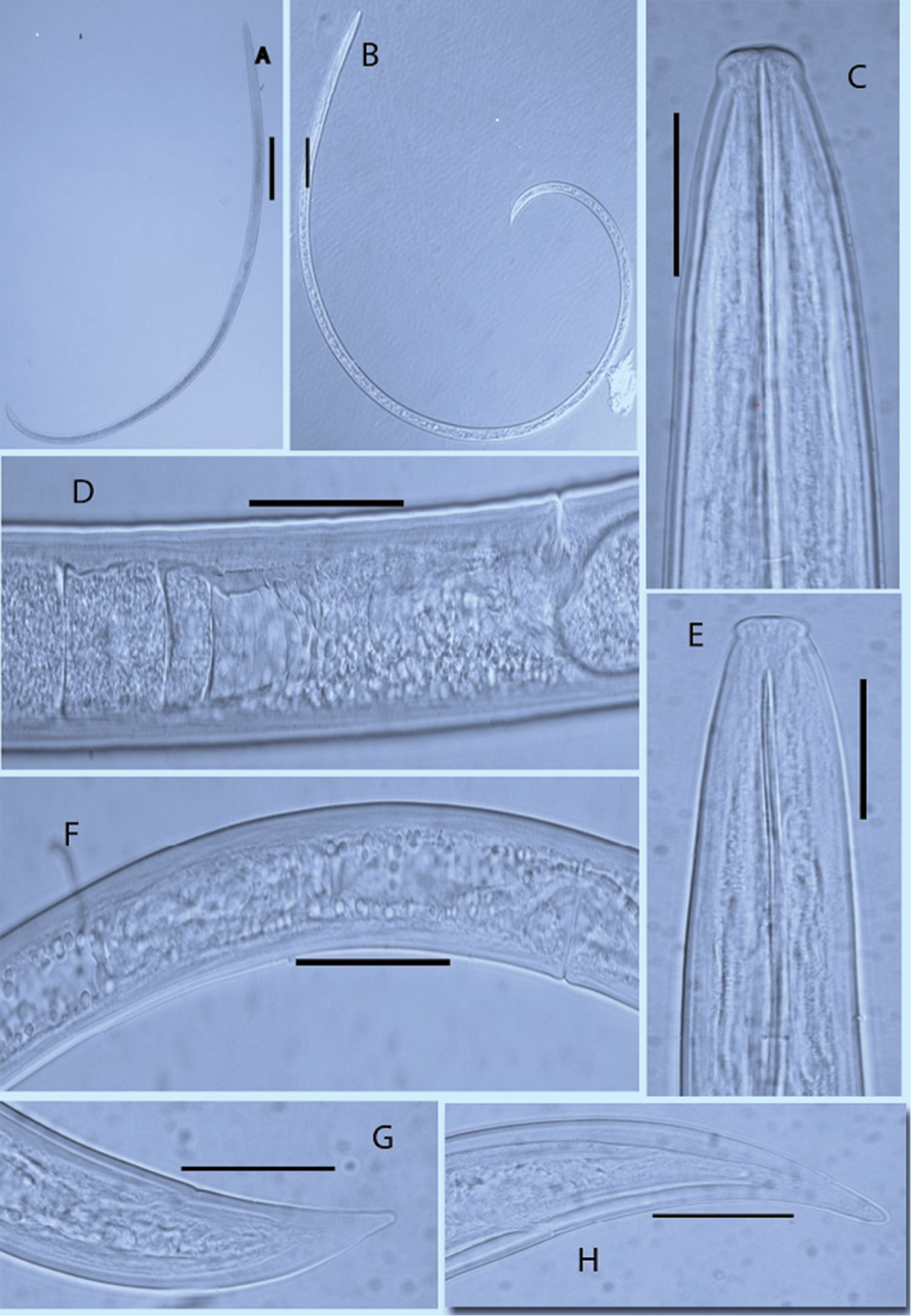
Photomicrographs of X. elongatum and X. pachtaicum. A, C, D, H Body habitus, head region, entire female reproductive part, tail region of X. elongatum respectively B, E, F, G Body habitus, head region; female reproductive part and tail part of X. pachtaicum respectively. A = 250 µm; B = 100 µm; D, F = 50 µm; C, E, G, H = 25 µm |