|
||
|
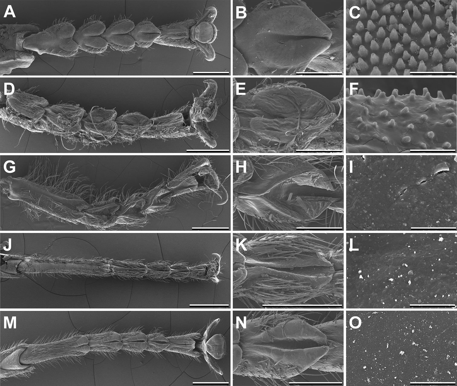
Scanning electron micrographs of the tarsal morphology of different Prisopodinae species. From left to right: Overview; Fourth euplantula; Adhesive microstructure. Scale bars: 1 mm; 300 μm; 5 μm. Melophasma antillarum, female (A–C). Paraprisopus merismus, female (D–F). Prisopus horstokkii, female (G–I). Dinelytron grylloides, female (J–L). Damasippus sp., female (M–O). |